- PDB-4l8o: Crystal structure of a bile-acid 7-alpha dehydratase (CLOHYLEM_06... -
+
Open data
ID or keywords:
Loading...
-
Basic information
Entry
Database: PDB / ID: 4l8o
Title
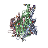
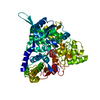
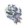
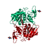
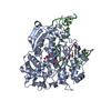
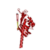
Crystal structure of a bile-acid 7-alpha dehydratase (CLOHYLEM_06634) from Clostridium hylemonae DSM 15053 at 2.20 A resolution
Components
Bile acid 7a-dehydratase, BaiE
Keywords
LYASE / SnoaL-like domain / PF13577 family protein / Structural Genomics / Joint Center for Structural Genomics / JCSG / Protein Structure Initiative / PSI-BIOLOGY
Resolution: 2.2→28.575 Å / Cor.coef. Fo:Fc: 0.942 / Cor.coef. Fo:Fc free: 0.932 / Occupancy max: 1 / Occupancy min: 0.33 / SU B: 9.914 / SU ML: 0.125 / Cross valid method: THROUGHOUT / σ(F): 0 / ESU R: 0.229 / ESU R Free: 0.184 / Stereochemistry target values: MAXIMUM LIKELIHOOD Details: 1. HYDROGENS HAVE BEEN ADDED IN THE RIDING POSITIONS. 2. ATOM RECORDS CONTAIN SUM OF TLS AND RESIDUAL B FACTORS. 3. ANISOU RECORDS CONTAIN SUM OF TLS AND RESIDUAL U FACTORS. 4. WATERS WERE ...Details: 1. HYDROGENS HAVE BEEN ADDED IN THE RIDING POSITIONS. 2. ATOM RECORDS CONTAIN SUM OF TLS AND RESIDUAL B FACTORS. 3. ANISOU RECORDS CONTAIN SUM OF TLS AND RESIDUAL U FACTORS. 4. WATERS WERE EXCLUDED FROM AUTOMATIC TLS ASSIGNMENT. 5. COBALT (CO+2) IS MODELED BASED ON A PEAK IN THE ANOMALOUS DIFFERENCE FOURIER AND ITS PRESCIENCE IN THE CRYSTALLIZATION CONDITIONS. 6. THE MODELED ETHYLENE GLYCOL (EDO), SULFATE (SO4) AND SODIUM IONS (NA) ARE PRESENT IN CRYO / CRYSTALLIZATION / PROTEIN BUFFER SOLUTIONS. 7. RESIDUES IN THE LOOP FROM 55-57 ALONG WITH PART OF THE NEIGHBORING RESIDUES 54 AND 58 HAVE BEEN LEFT UNMODELED. THIS LOOP IS NEAR A CRYSTALLOGRAPHIC 3-FOLD AXIS AND IT WAS DIFFICULT TO BUILD THE MULTIPLE CONFORMATIONS REQUIRED TO FIT THE DENSITY AND AVOID A SYMMETRY CLASH. 8. THE SCATTERING FACTORS FOR SODIUM, SULFUR, AND COBALT ATOMS WERE ADJUSTED BY REFMAC 5.7.0032 TO ACCOUNT FOR ANOMALOUS DISPERSION BASED ON THE WAVELENGTH 1.000 A (NA F'=0.06, S F'=0.18, CO F'=0.15). THE CROMER MANN VALUES LISTED IN THE CIF VERSION OF THE FILE INCLUDE THIS CORRECTION.
Rfactor
Num. reflection
% reflection
Selection details
Rfree
0.225
568
4.8 %
RANDOM
Rwork
0.1942
-
-
-
obs
0.1958
11786
94.73 %
-
Solvent computation
Ion probe radii: 0.8 Å / Shrinkage radii: 0.8 Å / VDW probe radii: 1.2 Å / Solvent model: BABINET MODEL WITH MASK
In the structure databanks used in Yorodumi, some data are registered as the other names, "COVID-19 virus" and "2019-nCoV". Here are the details of the virus and the list of structure data.
Jan 31, 2019. EMDB accession codes are about to change! (news from PDBe EMDB page)
EMDB accession codes are about to change! (news from PDBe EMDB page)
The allocation of 4 digits for EMDB accession codes will soon come to an end. Whilst these codes will remain in use, new EMDB accession codes will include an additional digit and will expand incrementally as the available range of codes is exhausted. The current 4-digit format prefixed with “EMD-” (i.e. EMD-XXXX) will advance to a 5-digit format (i.e. EMD-XXXXX), and so on. It is currently estimated that the 4-digit codes will be depleted around Spring 2019, at which point the 5-digit format will come into force.
The EM Navigator/Yorodumi systems omit the EMD- prefix.
Related info.:Q: What is EMD? / ID/Accession-code notation in Yorodumi/EM Navigator
Yorodumi is a browser for structure data from EMDB, PDB, SASBDB, etc.
This page is also the successor to EM Navigator detail page, and also detail information page/front-end page for Omokage search.
The word "yorodu" (or yorozu) is an old Japanese word meaning "ten thousand". "mi" (miru) is to see.
Related info.:EMDB / PDB / SASBDB / Comparison of 3 databanks / Yorodumi Search / Aug 31, 2016. New EM Navigator & Yorodumi / Yorodumi Papers / Jmol/JSmol / Function and homology information / Changes in new EM Navigator and Yorodumi
 Movie
Movie Controller

 Yorodumi
 Open data
 Basic information
 Components
 Keywords
 Function and homology information
 Clostridium hylemonae DSM 15053 (bacteria)
 X-RAY DIFFRACTION /
  Authors
 Citation
 Structure visualization
 Downloads & links
 Other downloads
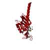


 PDBj
PDBj

 Assembly
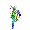


 Mass: 58.933 Da / Num. of mol.: 1 / Source method: obtained synthetically / Formula: Co
 Mass: 22.990 Da / Num. of mol.: 1 / Source method: obtained synthetically / Formula: Na
 Mass: 96.063 Da / Num. of mol.: 1 / Source method: obtained synthetically / Formula: SO4
 Mass: 62.068 Da / Num. of mol.: 4 / Source method: obtained synthetically / Formula: C2H6O2
 Sample preparation
 / Beamline: 8.2.2 / Wavelength: 0.999941
  Processing