 Movie
Movie Controller


+ Open data

- Basic information
| Entry | Database: PDB / ID: 1nbc | ||||||
|---|---|---|---|---|---|---|---|
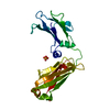
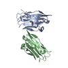
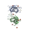
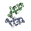
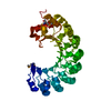
| Title | BACTERIAL TYPE 3A CELLULOSE-BINDING DOMAIN | ||||||
 Components Components | CELLULOSOMAL SCAFFOLDING PROTEIN A | ||||||
 Keywords Keywords | CELLULOSE DEGRADATION / CELLULOSE-BINDING DOMAIN / CELLULOSOME / SCAFOLDIN | ||||||
| Function / homology |  Function and homology information Function and homology informationcellulose binding / hydrolase activity, hydrolyzing O-glycosyl compounds / cellulose catabolic process / cell wall organization / extracellular region Similarity search - Function | ||||||
| Biological species |  Clostridium thermocellum (bacteria) Clostridium thermocellum (bacteria) | ||||||
| Method |  X-RAY DIFFRACTION / MIRAS / Resolution: 1.75 Å X-RAY DIFFRACTION / MIRAS / Resolution: 1.75 Å | ||||||
 Authors Authors | Tormo, J. / Lamed, R. / Steitz, T.A. | ||||||
 Citation Citation | Journal: EMBO J. / Year: 1996 Title: Crystal structure of a bacterial family-III cellulose-binding domain: a general mechanism for attachment to cellulose. Authors: Tormo, J. / Lamed, R. / Chirino, A.J. / Morag, E. / Bayer, E.A. / Shoham, Y. / Steitz, T.A. #1: Journal: Appl.Environ.Microbiol. / Year: 1995Title: Expression, Purification, and Characterization of the Cellulose-Binding Domain of the Scaffoldin Subunit from the Cellulosome of Clostridium Thermocellum Authors: Morag, E. / Lapidot, A. / Govorko, D. / Lamed, R. / Wilchek, M. / Bayer, E.A. / Shoham, Y. #2: Journal: J.Mol.Biol. / Year: 1994Title: Crystallization and Preliminary X-Ray Analysis of the Major Cellulose-Binding Domain of the Cellulosome from Clostridium Thermocellum Authors: Lamed, R. / Tormo, J. / Chirino, A.J. / Morag, E. / Bayer, E.A. | ||||||
| History |
|
- Structure visualization
| Structure viewer | Molecule: MolmilJmol/JSmol |
|---|
- Downloads & links
-Download
| PDBx/mmCIF format | 1nbc.cif.gz | 78.7 KB | Display | PDBx/mmCIF format |
|---|---|---|---|---|
| PDB format | pdb1nbc.ent.gz | 58.4 KB | Display | PDB format |
| PDBx/mmJSON format | 1nbc.json.gz | Tree view | PDBx/mmJSON format | |
| Others |  Other downloads Other downloads |
-Validation report
| Arichive directory | https://data.pdbj.org/pub/pdb/validation_reports/nb/1nbcftp://data.pdbj.org/pub/pdb/validation_reports/nb/1nbc | HTTPS FTP |
|---|
-Related structure data
| Similar structure data |
|---|
-Links
 PDBj
PDBj
- Assembly
| Deposited unit | 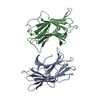
| ||||||||
|---|---|---|---|---|---|---|---|---|---|
| 1 |
| ||||||||
| Unit cell |
| ||||||||
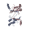
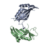
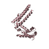
| Noncrystallographic symmetry (NCS) | NCS oper: (Code: given Matrix: (-0.999769, -0.006031, -0.020615), Vector: Details | THE ASYMMETRIC UNIT OF THE CRYSTAL CONTAINS TWO COPIES OF THE PROTEIN MOLECULE. CHAIN IDENTIFIER A: PROTOMER 1, CHAIN IDENTIFIER B: PROTOMER 2. | |
-Components
| #1: Protein | Mass: 17228.727 Da / Num. of mol.: 2 / Fragment: CELLULOSE-BINDING DOMAIN Source method: isolated from a genetically manipulated source Source: (gene. exp.) Clostridium thermocellum (bacteria) / Strain: YS / Gene: CIPB / Plasmid: PCBD / Gene (production host): CIPB / Production host: #2: Chemical |   Mass: 40.078 Da / Num. of mol.: 2 / Source method: obtained synthetically / Formula: Ca Mass: 40.078 Da / Num. of mol.: 2 / Source method: obtained synthetically / Formula: Ca#3: Water | ChemComp-HOH / |  Mass: 18.015 Da / Num. of mol.: 280 / Source method: isolated from a natural source / Formula: H2O Mass: 18.015 Da / Num. of mol.: 280 / Source method: isolated from a natural source / Formula: H2O |
|---|
-Experimental details
-Experiment
| Experiment | Method: X-RAY DIFFRACTION |
|---|
- Sample preparation
| Crystal | Density Matthews: 2.1 Å3/Da / Density % sol: 41 % | ||||||||||||||||||||||||||||||||||||||||
|---|---|---|---|---|---|---|---|---|---|---|---|---|---|---|---|---|---|---|---|---|---|---|---|---|---|---|---|---|---|---|---|---|---|---|---|---|---|---|---|---|---|
| Crystal grow | *PLUS Temperature: 12 ℃ / pH: 6.5 / Method: unknown | ||||||||||||||||||||||||||||||||||||||||
| Components of the solutions | *PLUS
|
-Data collection
| Diffraction | Mean temperature: 293 K |
|---|---|
| Diffraction source | Wavelength: 1.5418 |
| Detector | Type: RIGAKU / Detector: IMAGE PLATE / Details: YALE MIRRORS |
| Radiation | Monochromatic (M) / Laue (L): M / Scattering type: x-ray |
| Radiation wavelength | Wavelength: 1.5418 Å / Relative weight: 1 |
| Reflection | Resolution: 1.75→10 Å / Num. obs: 26267 / % possible obs: 87.4 % / Redundancy: 4.3 % / Biso Wilson estimate: 31.4 Å2 / Rmerge(I) obs: 0.082 |
| Reflection | *PLUS Num. measured all: 113902 |
| Reflection shell | *PLUS Highest resolution: 1.75 Å / Lowest resolution: 7.83 Å / % possible obs: 51 % |
- Processing
| Software |
| ||||||||||||||||||||||||||||||||||||||||||||||||||||||||||||||||||||||||||||||||
|---|---|---|---|---|---|---|---|---|---|---|---|---|---|---|---|---|---|---|---|---|---|---|---|---|---|---|---|---|---|---|---|---|---|---|---|---|---|---|---|---|---|---|---|---|---|---|---|---|---|---|---|---|---|---|---|---|---|---|---|---|---|---|---|---|---|---|---|---|---|---|---|---|---|---|---|---|---|---|---|---|---|
| Refinement | Method to determine structure: MIRAS / Resolution: 1.75→10 Å / σ(F): 2
| ||||||||||||||||||||||||||||||||||||||||||||||||||||||||||||||||||||||||||||||||
| Displacement parameters | Biso mean: 29.6 Å2
| ||||||||||||||||||||||||||||||||||||||||||||||||||||||||||||||||||||||||||||||||
| Refine analyze | Luzzati coordinate error obs: 0.23 Å / Luzzati d res low obs: 5 Å / Luzzati sigma a obs: 0.22 Å | ||||||||||||||||||||||||||||||||||||||||||||||||||||||||||||||||||||||||||||||||
| Refinement step | Cycle: LAST / Resolution: 1.75→10 Å
| ||||||||||||||||||||||||||||||||||||||||||||||||||||||||||||||||||||||||||||||||
| Refine LS restraints |
| ||||||||||||||||||||||||||||||||||||||||||||||||||||||||||||||||||||||||||||||||
| Xplor file |
| ||||||||||||||||||||||||||||||||||||||||||||||||||||||||||||||||||||||||||||||||
| Software | *PLUS Name: X-PLOR / Version: 3.1 / Classification: refinement | ||||||||||||||||||||||||||||||||||||||||||||||||||||||||||||||||||||||||||||||||
| Refinement | *PLUS Rfactor Rfree: 0.25 | ||||||||||||||||||||||||||||||||||||||||||||||||||||||||||||||||||||||||||||||||
| Solvent computation | *PLUS | ||||||||||||||||||||||||||||||||||||||||||||||||||||||||||||||||||||||||||||||||
| Displacement parameters | *PLUS | ||||||||||||||||||||||||||||||||||||||||||||||||||||||||||||||||||||||||||||||||
| Refine LS restraints | *PLUS
|
