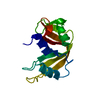
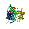
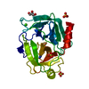
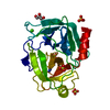
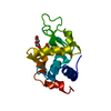
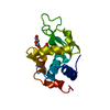
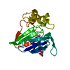
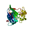
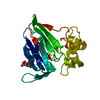
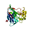
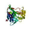
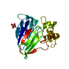
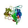
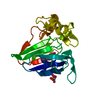
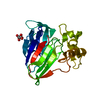
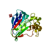
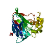
SHEET THE SHEET STRUCTURE OF THIS MOLECULE IS BIFURCATED. IN ORDER TO REPRESENT THIS FEATURE IN ... SHEET THE SHEET STRUCTURE OF THIS MOLECULE IS BIFURCATED. IN ORDER TO REPRESENT THIS FEATURE IN THE SHEET RECORDS BELOW, TWO SHEETS ARE DEFINED.
Type: ADSC CCD / Detector: CCD / Date: Dec 18, 2004 / Details: BENT MIRROR
Radiation
Monochromator: SI(111) / Protocol: SINGLE WAVELENGTH / Monochromatic (M) / Laue (L): M / Scattering type: x-ray
Radiation wavelength
Wavelength: 0.9392 Å / Relative weight: 1
Reflection
Resolution: 1.4→50 Å / Num. obs: 93386 / % possible obs: 97.6 % / Observed criterion σ(I): -3 / Redundancy: 2.5 % / Rmerge(I) obs: 0.04 / Net I/σ(I): 15.8
Reflection shell
Resolution: 1.4→1.49 Å / Redundancy: 2.5 % / Rmerge(I) obs: 0.26 / Mean I/σ(I) obs: 3.81 / % possible all: 94.8
-
Processing
Software
Name
Version
Classification
XDS
datareduction
XSCALE
datascaling
SHELXD
phasing
SHELXE
phasing
REFMAC
5.2.0005
refinement
Refinement
Method to determine structure: OTHER / Resolution: 1.4→45 Å / Cor.coef. Fo:Fc: 0.974 / Cor.coef. Fo:Fc free: 0.965 / SU B: 1.279 / SU ML: 0.023 / Cross valid method: THROUGHOUT / ESU R: 0.045 / ESU R Free: 0.045 / Stereochemistry target values: MAXIMUM LIKELIHOOD / Details: HYDROGENS HAVE BEEN ADDED IN THE RIDING POSITIONS
Rfactor
Num. reflection
% reflection
Selection details
Rfree
0.155
2538
5.1 %
RANDOM
Rwork
0.126
-
-
-
obs
0.127
47378
97.9 %
-
Solvent computation
Ion probe radii: 0.8 Å / Shrinkage radii: 0.8 Å / VDW probe radii: 1.2 Å / Solvent model: BABINET MODEL WITH MASK
 Movie
Movie Controller


 Open data
 Basic information
 Components
 Keywords
 Function and homology information
 THAUMATOCOCCUS DANIELLII (katemfe)
 X-RAY DIFFRACTION /
  Authors
 Citation
 Structure visualization
 Downloads & links
 Other downloads
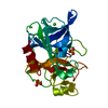


 PDBj
PDBj

 Assembly

 Mass: 150.087 Da / Num. of mol.: 1 / Source method: obtained synthetically / Formula: C4H6O6
 Mass: 18.015 Da / Num. of mol.: 276 / Source method: isolated from a natural source / Formula: H2O
 Sample preparation
 / Beamline: ID14-4 / Wavelength: 0.9392
  Processing