 Movie
Movie Controller

[English] 日本語
 Yorodumi
- PDB-2ahs: Crystal Structure of the Catalytic Domain of Human Tyrosine Recep... -
+ Open data

- Basic information
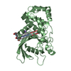
| Entry | Database: PDB / ID: 2ahs | ||||||
|---|---|---|---|---|---|---|---|
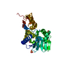
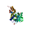
| Title | Crystal Structure of the Catalytic Domain of Human Tyrosine Receptor Phosphatase Beta | ||||||
 Components Components | Receptor-type tyrosine-protein phosphatase beta | ||||||
 Keywords Keywords | HYDROLASE / Tyrosine Receptor Phosphatase / Beta / Human / Structural Genomics / Structural Genomics Consortium / SGC | ||||||
| Function / homology |  Function and homology information Function and homology informationtransmembrane receptor protein tyrosine kinase inhibitor activity / glial cell migration / transmembrane receptor protein tyrosine phosphatase activity / dephosphorylation / phosphate-containing compound metabolic process / protein dephosphorylation / tertiary granule membrane / specific granule membrane / protein-tyrosine-phosphatase / osteoblast differentiation ...transmembrane receptor protein tyrosine kinase inhibitor activity / glial cell migration / transmembrane receptor protein tyrosine phosphatase activity / dephosphorylation / phosphate-containing compound metabolic process / protein dephosphorylation / tertiary granule membrane / specific granule membrane / protein-tyrosine-phosphatase / osteoblast differentiation / angiogenesis / signaling receptor complex / cadherin binding / Neutrophil degranulation / plasma membrane Similarity search - Function | ||||||
| Biological species |  Homo sapiens (human) Homo sapiens (human) | ||||||
| Method |  X-RAY DIFFRACTION / SYNCHROTRON / MOLECULAR REPLACEMENT / Resolution: 2.1 Å X-RAY DIFFRACTION / SYNCHROTRON / MOLECULAR REPLACEMENT / Resolution: 2.1 Å | ||||||
 Authors Authors | Ugochukwu, E. / Eswaran, J. / Barr, A. / Gileadi, O. / Sobott, F. / Burgess, N. / Ball, L. / Bray, J. / von Delft, F. / Debreczeni, J. ...Ugochukwu, E. / Eswaran, J. / Barr, A. / Gileadi, O. / Sobott, F. / Burgess, N. / Ball, L. / Bray, J. / von Delft, F. / Debreczeni, J. / Bunkoczi, G. / Turnbull, A. / Das, S. / Weigelt, J. / Edwards, A. / Arrowsmith, C. / Sundstrom, M. / Knapp, S. / Structural Genomics Consortium (SGC) | ||||||
 Citation Citation | Journal: Cell(Cambridge,Mass.) / Year: 2009 Title: Large-scale structural analysis of the classical human protein tyrosine phosphatome. Authors: Barr, A.J. / Ugochukwu, E. / Lee, W.H. / King, O.N. / Filippakopoulos, P. / Alfano, I. / Savitsky, P. / Burgess-Brown, N.A. / Muller, S. / Knapp, S. | ||||||
| History |
|
- Structure visualization
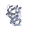
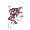
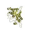
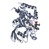
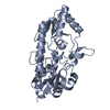
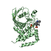
| Structure viewer | Molecule: MolmilJmol/JSmol |
|---|
- Downloads & links
-Download
| PDBx/mmCIF format | 2ahs.cif.gz | 139.8 KB | Display | PDBx/mmCIF format |
|---|---|---|---|---|
| PDB format | pdb2ahs.ent.gz | 108 KB | Display | PDB format |
| PDBx/mmJSON format | 2ahs.json.gz | Tree view | PDBx/mmJSON format | |
| Others |  Other downloads Other downloads |
-Validation report
| Arichive directory | https://data.pdbj.org/pub/pdb/validation_reports/ah/2ahsftp://data.pdbj.org/pub/pdb/validation_reports/ah/2ahs | HTTPS FTP |
|---|
-Related structure data
| Related structure data |  2b49C  2cfvC 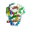 2cjzC  2gjtC  2h4vC  2i75C  2jjdC  2nlkC  2nz6C  2oc3C  2ooqC  2p6xC  2pa5C  2qepC  3b7oC  1rpmS S: Starting model for refinement C: citing same article ( |
|---|---|
| Similar structure data |

-Links
 PDBj
PDBj


- Assembly
| Deposited unit | 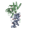
| |||||||||||||||||||||||||||||||||||||||||||||||||||||||||||||||||||||||||||||||||||||||||||||||||||||||||||||||||||||||||||||||||||||||||||||||||||||||||||||||||||||||||||||||||||||||||||||||||||||||||||||||||
|---|---|---|---|---|---|---|---|---|---|---|---|---|---|---|---|---|---|---|---|---|---|---|---|---|---|---|---|---|---|---|---|---|---|---|---|---|---|---|---|---|---|---|---|---|---|---|---|---|---|---|---|---|---|---|---|---|---|---|---|---|---|---|---|---|---|---|---|---|---|---|---|---|---|---|---|---|---|---|---|---|---|---|---|---|---|---|---|---|---|---|---|---|---|---|---|---|---|---|---|---|---|---|---|---|---|---|---|---|---|---|---|---|---|---|---|---|---|---|---|---|---|---|---|---|---|---|---|---|---|---|---|---|---|---|---|---|---|---|---|---|---|---|---|---|---|---|---|---|---|---|---|---|---|---|---|---|---|---|---|---|---|---|---|---|---|---|---|---|---|---|---|---|---|---|---|---|---|---|---|---|---|---|---|---|---|---|---|---|---|---|---|---|---|---|---|---|---|---|---|---|---|---|---|---|---|---|---|---|---|---|
| 1 | 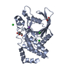
| |||||||||||||||||||||||||||||||||||||||||||||||||||||||||||||||||||||||||||||||||||||||||||||||||||||||||||||||||||||||||||||||||||||||||||||||||||||||||||||||||||||||||||||||||||||||||||||||||||||||||||||||||
| 2 | 
| |||||||||||||||||||||||||||||||||||||||||||||||||||||||||||||||||||||||||||||||||||||||||||||||||||||||||||||||||||||||||||||||||||||||||||||||||||||||||||||||||||||||||||||||||||||||||||||||||||||||||||||||||
| Unit cell |
| |||||||||||||||||||||||||||||||||||||||||||||||||||||||||||||||||||||||||||||||||||||||||||||||||||||||||||||||||||||||||||||||||||||||||||||||||||||||||||||||||||||||||||||||||||||||||||||||||||||||||||||||||
| Noncrystallographic symmetry (NCS) | NCS domain:
NCS domain segments: Ens-ID: 1
|