| Entry | Database: PDB / ID: 2vc4
|
|---|
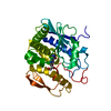
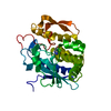
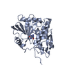
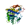
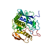
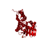
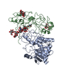
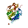
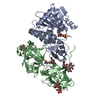
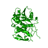
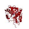
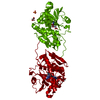
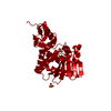
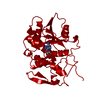
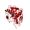
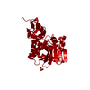
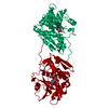
| Title | Ricin A-Chain (Recombinant) E177D Mutant |
|---|
 Components Components | RICIN A CHAIN |
|---|
 Keywords Keywords | HYDROLASE / GLYCOPROTEIN / PLANT DEFENSE / PROTEIN SYNTHESIS INHIBITOR / TOXIN / LECTIN / GLYCOSIDASE |
|---|
| Function / homology |  Function and homology information Function and homology information
rRNA N-glycosylase / rRNA N-glycosylase activity / AMP binding / defense response / toxin activity / carbohydrate binding / killing of cells of another organism / negative regulation of translationSimilarity search - Function Ricin (A Subunit), domain 2 / Ricin (A Subunit), domain 2 / Ricin (A subunit); domain 1 / Ricin (A subunit), domain 1 / Ricin-type beta-trefoil lectin domain / Ribosome-inactivating protein conserved site / Shiga/ricin ribosomal inactivating toxins active site signature. / Ribosome-inactivating protein type 1/2 / Ribosome-inactivating protein / Ribosome-inactivating protein, subdomain 1 ...Ricin (A Subunit), domain 2 / Ricin (A Subunit), domain 2 / Ricin (A subunit); domain 1 / Ricin (A subunit), domain 1 / Ricin-type beta-trefoil lectin domain / Ribosome-inactivating protein conserved site / Shiga/ricin ribosomal inactivating toxins active site signature. / Ribosome-inactivating protein type 1/2 / Ribosome-inactivating protein / Ribosome-inactivating protein, subdomain 1 / Ribosome-inactivating protein, subdomain 2 / Ribosome-inactivating protein superfamily / Ribosome inactivating protein / Ricin-type beta-trefoil / Lectin domain of ricin B chain profile. / Ricin B, lectin domain / Ricin B-like lectins / Few Secondary Structures / Irregular / 3-Layer(aba) Sandwich / Alpha BetaSimilarity search - Domain/homology |
|---|
| Biological species |  RICINUS COMMUNIS (castor bean) RICINUS COMMUNIS (castor bean) |
|---|
| Method |  X-RAY DIFFRACTION / SYNCHROTRON / MOLECULAR REPLACEMENT / Resolution: 1.39 Å X-RAY DIFFRACTION / SYNCHROTRON / MOLECULAR REPLACEMENT / Resolution: 1.39 Å |
|---|
 Authors Authors | Marsden, C.J. / Fulop, V. |
|---|
 Citation Citation | Journal: FEBS J. / Year: 2007
Title: The Isolation and Characterisation of Temperature-Dependent Ricin a Chain Molecules in Saccharomyces Cerevisiae
Authors: Allen, S.C.H. / Moore, K.A.H. / Marsden, C.J. / Fulop, V. / Moffat, K.G. / Lord, J.M. / Ladds, G. / Roberts, L.M. |
|---|
| History | | Deposition | Sep 18, 2007 | Deposition site: PDBE / Processing site: PDBE |
|---|
| Revision 1.0 | Oct 16, 2007 | Provider: repository / Type: Initial release |
|---|
| Revision 1.1 | Jul 13, 2011 | Group: Advisory / Version format compliance |
|---|
| Revision 1.2 | Jan 17, 2018 | Group: Data collection / Category: diffrn_source / Item: _diffrn_source.pdbx_synchrotron_site |
|---|
| Revision 1.3 | Dec 13, 2023 | Group: Data collection / Database references ...Data collection / Database references / Other / Refinement description
Category: chem_comp_atom / chem_comp_bond ...chem_comp_atom / chem_comp_bond / database_2 / pdbx_database_status / pdbx_initial_refinement_model
Item: _database_2.pdbx_DOI / _database_2.pdbx_database_accession / _pdbx_database_status.status_code_sf |
|---|
|
|---|
 Movie
Movie Controller


 Open data
 Basic information
 Structure visualization
 Downloads & links
 Other downloads


 PDBj
PDBj

 Assembly


 Mass: 92.094 Da / Num. of mol.: 1 / Source method: obtained synthetically / Formula: C3H8O3
 Mass: 96.063 Da / Num. of mol.: 2 / Source method: obtained synthetically / Formula: SO4
 Mass: 18.015 Da / Num. of mol.: 404 / Source method: isolated from a natural source / Formula: H2O
 Sample preparation
 / Beamline: I711 / Wavelength: 1.0213
  Processing