 Movie
Movie Controller


+ Open data

- Basic information
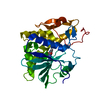
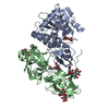
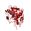
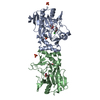
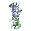
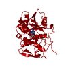
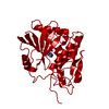
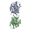
| Entry | Database: PDB / ID: 1uq4 | ||||||
|---|---|---|---|---|---|---|---|
| Title | RICIN A-CHAIN (RECOMBINANT) R213D MUTANT | ||||||
 Components Components | RICIN | ||||||
 Keywords Keywords | HYDROLASE / GLYCOSIDASE / TOXIN / GLYCOPROTEIN | ||||||
| Function / homology |  Function and homology information Function and homology informationrRNA N-glycosylase / rRNA N-glycosylase activity / AMP binding / defense response / toxin activity / carbohydrate binding / killing of cells of another organism / negative regulation of translation Similarity search - Function | ||||||
| Biological species |  RICINUS COMMUNIS (castor bean) RICINUS COMMUNIS (castor bean) | ||||||
| Method |  X-RAY DIFFRACTION / MOLECULAR REPLACEMENT / Resolution: 1.9 Å X-RAY DIFFRACTION / MOLECULAR REPLACEMENT / Resolution: 1.9 Å | ||||||
 Authors Authors | Marsden, C.J. / Fulop, V. | ||||||
 Citation Citation | Journal: Eur.J.Biochem. / Year: 2004 Title: The Effect of Mutations Surrounding and within the Active Site on the Catalytic Activity of Ricin a Chain Authors: Marsden, C.J. / Fulop, V. / Day, P. / Lord, J.M. #1: Journal: J.Mol.Biol. / Year: 1994Title: X-Ray Structure of Recombinant Ricin A-Chain at 1.8 A Resolution Authors: Weston, S.A. / Tucker, A.D. / Thatcher, D.R. / Derbyshire, D.J. / Pauptit, R.A. | ||||||
| History |
|
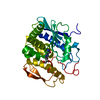
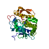
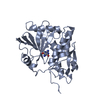
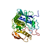
- Structure visualization
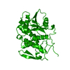
| Structure viewer | Molecule: MolmilJmol/JSmol |
|---|
- Downloads & links
-Download
| PDBx/mmCIF format | 1uq4.cif.gz | 75.2 KB | Display | PDBx/mmCIF format |
|---|---|---|---|---|
| PDB format | pdb1uq4.ent.gz | 55.2 KB | Display | PDB format |
| PDBx/mmJSON format | 1uq4.json.gz | Tree view | PDBx/mmJSON format | |
| Others |  Other downloads Other downloads |
-Validation report
| Arichive directory | https://data.pdbj.org/pub/pdb/validation_reports/uq/1uq4ftp://data.pdbj.org/pub/pdb/validation_reports/uq/1uq4 | HTTPS FTP |
|---|
-Related structure data
| Related structure data | 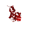 1uq5C  1iftS S: Starting model for refinement C: citing same article ( |
|---|---|
| Similar structure data |


-Links
 PDBj
PDBj

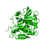
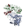
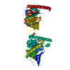
- Assembly
| Deposited unit | 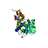
| ||||||||||||
|---|---|---|---|---|---|---|---|---|---|---|---|---|---|
| 1 | 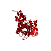
| ||||||||||||
| Unit cell |
| ||||||||||||
| Components on special symmetry positions |
| ||||||||||||
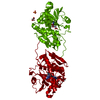
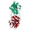
| Details | THIS DIMER IS BECAUSE OF CRYSTAL PACKING BUT NONETHELESSGIVES AN INTERESTING RESULT |
-Components
| #1: Protein | Mass: 29408.029 Da / Num. of mol.: 1 / Fragment: A CHAIN, RESIDUES 40-302 / Mutation: YES Source method: isolated from a genetically manipulated source Source: (gene. exp.) RICINUS COMMUNIS (castor bean) / Production host:  | ||||
|---|---|---|---|---|---|
| #2: Chemical |   Mass: 96.063 Da / Num. of mol.: 2 / Source method: obtained synthetically / Formula: SO4 Mass: 96.063 Da / Num. of mol.: 2 / Source method: obtained synthetically / Formula: SO4#3: Water | ChemComp-HOH / |  Mass: 18.015 Da / Num. of mol.: 406 / Source method: isolated from a natural source / Formula: H2O Mass: 18.015 Da / Num. of mol.: 406 / Source method: isolated from a natural source / Formula: H2OCompound details | ENGINEERED | |
-Experimental details
-Experiment
| Experiment | Method: X-RAY DIFFRACTION / Number of used crystals: 1 |
|---|
- Sample preparation
| Crystal | Density Matthews: 2.7 Å3/Da / Density % sol: 48 % | |||||||||||||||||||||||||||||||||||||||||||||
|---|---|---|---|---|---|---|---|---|---|---|---|---|---|---|---|---|---|---|---|---|---|---|---|---|---|---|---|---|---|---|---|---|---|---|---|---|---|---|---|---|---|---|---|---|---|---|
| Crystal grow | pH: 4.2 / Details: SEE REFERENCE 1, pH 4.20 | |||||||||||||||||||||||||||||||||||||||||||||
| Crystal grow | *PLUS Temperature: 4 ℃ / pH: 8.9 / Method: vapor diffusion, hanging drop / Details: Weston, S.A., (1994) J. Mol. Biol., 244, 410. | |||||||||||||||||||||||||||||||||||||||||||||
| Components of the solutions | *PLUS
|
-Data collection
| Diffraction | Mean temperature: 100 K |
|---|---|
| Diffraction source | Source: ROTATING ANODE / Type: ENRAF-NONIUS FR591 / Wavelength: 1.5418 |
| Detector | Type: MAC Science DIP-2030 / Detector: IMAGE PLATE / Date: Nov 17, 2000 / Details: MIRRORS |
| Radiation | Protocol: SINGLE WAVELENGTH / Monochromatic (M) / Laue (L): M / Scattering type: x-ray |
| Radiation wavelength | Wavelength: 1.5418 Å / Relative weight: 1 |
| Reflection | Resolution: 1.9→28 Å / Num. obs: 53233 / % possible obs: 86 % / Observed criterion σ(I): -3 / Redundancy: 2.4 % / Biso Wilson estimate: 21.9 Å2 / Rmerge(I) obs: 0.038 / Net I/σ(I): 21.6 |
| Reflection shell | Resolution: 1.9→1.97 Å / Redundancy: 1.5 % / Rmerge(I) obs: 0.133 / Mean I/σ(I) obs: 4.7 / % possible all: 44 |
| Reflection | *PLUS Highest resolution: 1.9 Å / Lowest resolution: 28 Å / Num. obs: 22494 / % possible obs: 85.6 % / Num. measured all: 53233 / Rmerge(I) obs: 0.038 |
| Reflection shell | *PLUS Highest resolution: 1.9 Å / Lowest resolution: 1.949 Å / % possible obs: 43.6 % / Rmerge(I) obs: 0.133 / Mean I/σ(I) obs: 4.7 |
- Processing
| Software |
| ||||||||||||||||||||
|---|---|---|---|---|---|---|---|---|---|---|---|---|---|---|---|---|---|---|---|---|---|
| Refinement | Method to determine structure: MOLECULAR REPLACEMENT Starting model: PDB ENTRY 1IFT Resolution: 1.9→28 Å / SU B: 3.03 / SU ML: 0.089 / Cross valid method: THROUGHOUT / σ(F): 0 / ESU R: 0.145 / ESU R Free: 0.159
| ||||||||||||||||||||
| Displacement parameters | Biso mean: 24.8 Å2
| ||||||||||||||||||||
| Refinement step | Cycle: LAST / Resolution: 1.9→28 Å
| ||||||||||||||||||||
| Refinement | *PLUS % reflection Rfree: 4 % | ||||||||||||||||||||
| Solvent computation | *PLUS | ||||||||||||||||||||
| Displacement parameters | *PLUS | ||||||||||||||||||||
| Refine LS restraints | *PLUS
| ||||||||||||||||||||
| LS refinement shell | *PLUS Rfactor Rfree: 0.257 / Num. reflection Rfree: 24 / Rfactor Rwork: 0.154 / Num. reflection Rwork: 720 |
