 Movie
Movie Controller


+ Open data

- Basic information
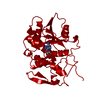
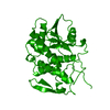
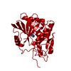
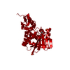
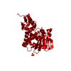
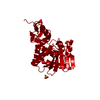
| Entry | Database: PDB / ID: 1obs | ||||||
|---|---|---|---|---|---|---|---|
| Title | STRUCTURE OF RICIN A CHAIN MUTANT | ||||||
 Components Components | RICIN A CHAIN | ||||||
 Keywords Keywords | GLYCOSIDASE / HYDROLASE / TOXIN / DUPLICATION / GLYCOPROTEIN / LECTIN | ||||||
| Function / homology |  Function and homology information Function and homology informationrRNA N-glycosylase / rRNA N-glycosylase activity / AMP binding / defense response / toxin activity / carbohydrate binding / killing of cells of another organism / negative regulation of translation Similarity search - Function | ||||||
| Biological species |  Ricinus communis (castor bean) Ricinus communis (castor bean) | ||||||
| Method |  X-RAY DIFFRACTION / Resolution: 2.2 Å X-RAY DIFFRACTION / Resolution: 2.2 Å | ||||||
 Authors Authors | Day, P.J. / Ernst, S.R. / Frankel, A.E. / Monzingo, A.F. / Pascal, J.M. / Svinth, M. / Robertus, J.D. | ||||||
 Citation Citation | Journal: Biochemistry / Year: 1996 Title: Structure and activity of an active site substitution of ricin A chain. Authors: Day, P.J. / Ernst, S.R. / Frankel, A.E. / Monzingo, A.F. / Pascal, J.M. / Molina-Svinth, M.C. / Robertus, J.D. #1: Journal: J.Mol.Biol. / Year: 1994Title: X-Ray Structure of Recombinant Ricin A-Chain at 1.8 A Resolution Authors: Weston, S.A. / Tucker, A.D. / Thatcher, D.R. / Derbyshire, D.J. / Pauptit, R.A. #2: Journal: Protein Sci. / Year: 1993Title: Structure of Recombinant Ricin a Chain at 2.3 A Authors: Mlsna, D. / Monzingo, A.F. / Katzin, B.J. / Ernst, S. / Robertus, J.D. | ||||||
| History |
|
- Structure visualization
| Structure viewer | Molecule: MolmilJmol/JSmol |
|---|
- Downloads & links
-Download
| PDBx/mmCIF format | 1obs.cif.gz | 57.3 KB | Display | PDBx/mmCIF format |
|---|---|---|---|---|
| PDB format | pdb1obs.ent.gz | 41 KB | Display | PDB format |
| PDBx/mmJSON format | 1obs.json.gz | Tree view | PDBx/mmJSON format | |
| Others |  Other downloads Other downloads |
-Validation report
| Arichive directory | https://data.pdbj.org/pub/pdb/validation_reports/ob/1obsftp://data.pdbj.org/pub/pdb/validation_reports/ob/1obs | HTTPS FTP |
|---|
-Related structure data


-Links
 PDBj
PDBj

- Assembly
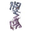
| Deposited unit | 
| ||||||||
|---|---|---|---|---|---|---|---|---|---|
| 1 | 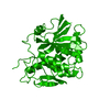
| ||||||||
| Unit cell |
|
-Components
| #1: Protein | Mass: 29917.711 Da / Num. of mol.: 1 / Mutation: R180H Source method: isolated from a genetically manipulated source Source: (gene. exp.) Ricinus communis (castor bean) / Plasmid: PUTA / Production host:  |
|---|---|
| #2: Water | ChemComp-HOH /  Mass: 18.015 Da / Num. of mol.: 87 / Source method: isolated from a natural source / Formula: H2O Mass: 18.015 Da / Num. of mol.: 87 / Source method: isolated from a natural source / Formula: H2O |
-Experimental details
-Experiment
| Experiment | Method: X-RAY DIFFRACTION |
|---|
- Sample preparation
| Crystal | Density Matthews: 2.79 Å3/Da / Density % sol: 55.93 % | ||||||||||||||||||||||||||||||||||||||||
|---|---|---|---|---|---|---|---|---|---|---|---|---|---|---|---|---|---|---|---|---|---|---|---|---|---|---|---|---|---|---|---|---|---|---|---|---|---|---|---|---|---|
| Crystal grow | *PLUS Temperature: 4 ℃ / pH: 6.5 / Method: vapor diffusion, sitting drop | ||||||||||||||||||||||||||||||||||||||||
| Components of the solutions | *PLUS
|
-Data collection
| Diffraction source | Wavelength: 1.5418 |
|---|---|
| Detector | Type: XUONG-HAMLIN MULTIWIRE / Detector: AREA DETECTOR / Date: Mar 6, 1995 |
| Radiation | Monochromatic (M) / Laue (L): M / Scattering type: x-ray |
| Radiation wavelength | Wavelength: 1.5418 Å / Relative weight: 1 |
| Reflection | Resolution: 2.1→50 Å / Num. obs: 18873 / % possible obs: 90.9 % / Observed criterion σ(I): 0 / Redundancy: 20 % / Rmerge(I) obs: 0.076 |
| Reflection | *PLUS Num. measured all: 379920 |
- Processing
| Software |
| ||||||||||||||||||||||||||||||||||||||||||||||||||||||||||||
|---|---|---|---|---|---|---|---|---|---|---|---|---|---|---|---|---|---|---|---|---|---|---|---|---|---|---|---|---|---|---|---|---|---|---|---|---|---|---|---|---|---|---|---|---|---|---|---|---|---|---|---|---|---|---|---|---|---|---|---|---|---|
| Refinement | Resolution: 2.2→10 Å / σ(F): 2 Details: SET OF IDEAL BOND LENGTHS AND ANGLES USED DURING REFINEMENT: RAMACHANDRAN ET AL. (1974), BIOCHIM. BIOPHYS. ACTA 359, 298. FINAL RMS COORD. SHIFT 0.49 ANGSTROMS
| ||||||||||||||||||||||||||||||||||||||||||||||||||||||||||||
| Refinement step | Cycle: LAST / Resolution: 2.2→10 Å
| ||||||||||||||||||||||||||||||||||||||||||||||||||||||||||||
| Refine LS restraints |
| ||||||||||||||||||||||||||||||||||||||||||||||||||||||||||||
| Software | *PLUS Name: X-PLOR / Version: 3.1 / Classification: refinement | ||||||||||||||||||||||||||||||||||||||||||||||||||||||||||||
| Refine LS restraints | *PLUS
|
