 Movie
Movie Controller

[English] 日本語
 Yorodumi
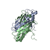
- PDB-3b7o: Crystal structure of the human tyrosine phosphatase SHP2 (PTPN11)... -
+ Open data

- Basic information
| Entry | Database: PDB / ID: 3b7o | ||||||
|---|---|---|---|---|---|---|---|
| Title | Crystal structure of the human tyrosine phosphatase SHP2 (PTPN11) with an accessible active site | ||||||
 Components Components | Tyrosine-protein phosphatase non-receptor type 11 | ||||||
 Keywords Keywords | HYDROLASE / SHP2 / PTPN11 / tyrosine phosphatase / Structural Genomics / Structural Genomics Consortium / SGC / Deafness / Disease mutation / Phosphorylation / Protein phosphatase / SH2 domain | ||||||
| Function / homology |  Function and homology information Function and homology informationnegative regulation of cortisol secretion / intestinal epithelial cell migration / microvillus organization / negative regulation of growth hormone secretion / atrioventricular canal development / genitalia development / STAT5 Activation / Co-inhibition by BTLA / Netrin mediated repulsion signals / negative regulation of neutrophil activation ...negative regulation of cortisol secretion / intestinal epithelial cell migration / microvillus organization / negative regulation of growth hormone secretion / atrioventricular canal development / genitalia development / STAT5 Activation / Co-inhibition by BTLA / Netrin mediated repulsion signals / negative regulation of neutrophil activation / cerebellar cortex formation / positive regulation of hormone secretion / regulation of protein export from nucleus / positive regulation of lipopolysaccharide-mediated signaling pathway / Interleukin-37 signaling / Signaling by Leptin / negative regulation of cell adhesion mediated by integrin / positive regulation of ossification / hormone metabolic process / MET activates PTPN11 / negative regulation of chondrocyte differentiation / Regulation of RUNX1 Expression and Activity / Signal regulatory protein family interactions / face morphogenesis / platelet formation / organ growth / ERBB signaling pathway / triglyceride metabolic process / megakaryocyte development / Interleukin-20 family signaling / Interleukin-6 signaling / Co-inhibition by CTLA4 / PI-3K cascade:FGFR3 / Platelet sensitization by LDL / STAT5 activation downstream of FLT3 ITD mutants / PI-3K cascade:FGFR2 / peptide hormone receptor binding / PI-3K cascade:FGFR4 / negative regulation of T cell activation / MAPK3 (ERK1) activation / negative regulation of type I interferon production / PI-3K cascade:FGFR1 / neurotrophin TRK receptor signaling pathway / regulation of type I interferon-mediated signaling pathway / platelet-derived growth factor receptor signaling pathway / MAPK1 (ERK2) activation / Prolactin receptor signaling / inner ear development / PECAM1 interactions / Bergmann glial cell differentiation / peptidyl-tyrosine dephosphorylation / non-membrane spanning protein tyrosine phosphatase activity / Regulation of IFNA/IFNB signaling / RET signaling / positive regulation of intracellular signal transduction / Interleukin-3, Interleukin-5 and GM-CSF signaling / fibroblast growth factor receptor signaling pathway / Co-inhibition by PD-1 / PI3K Cascade / ephrin receptor signaling pathway / positive regulation of insulin receptor signaling pathway / regulation of protein-containing complex assembly / Regulation of IFNG signaling / GAB1 signalosome / negative regulation of T cell receptor signaling pathway / Activated NTRK2 signals through FRS2 and FRS3 / negative regulation of T cell proliferation / GPVI-mediated activation cascade / T cell costimulation / Signaling by CSF3 (G-CSF) / FRS-mediated FGFR3 signaling / Signaling by FLT3 ITD and TKD mutants / phosphotyrosine residue binding / FRS-mediated FGFR2 signaling / FRS-mediated FGFR4 signaling / phosphoprotein phosphatase activity / FRS-mediated FGFR1 signaling / protein-tyrosine-phosphatase / Tie2 Signaling / hormone-mediated signaling pathway / FLT3 Signaling / positive regulation of mitotic cell cycle / protein tyrosine phosphatase activity / axonogenesis / cell adhesion molecule binding / positive regulation of interferon-beta production / Downstream signal transduction / homeostasis of number of cells within a tissue / DNA damage checkpoint signaling / cellular response to epidermal growth factor stimulus / protein tyrosine kinase binding / positive regulation of D-glucose import across plasma membrane / insulin receptor binding / integrin-mediated signaling pathway / Activation of IRF3, IRF7 mediated by TBK1, IKKε (IKBKE) / Negative regulation of FGFR3 signaling / Negative regulation of FGFR2 signaling / cellular response to mechanical stimulus / Negative regulation of FGFR4 signaling / Negative regulation of FGFR1 signaling Similarity search - Function | ||||||
| Biological species |  Homo sapiens (human) Homo sapiens (human) | ||||||
| Method |  X-RAY DIFFRACTION / SYNCHROTRON / MOLECULAR REPLACEMENT / Resolution: 1.6 Å X-RAY DIFFRACTION / SYNCHROTRON / MOLECULAR REPLACEMENT / Resolution: 1.6 Å | ||||||
 Authors Authors | Ugochukwu, E. / Barr, A. / Patel, A. / King, O. / Niesen, F. / Salah, E. / Savitsky, P. / Pilka, E.S. / Elkins, J. / Arrowsmith, C.H. ...Ugochukwu, E. / Barr, A. / Patel, A. / King, O. / Niesen, F. / Salah, E. / Savitsky, P. / Pilka, E.S. / Elkins, J. / Arrowsmith, C.H. / Weigelt, J. / Edwards, A.M. / von Delft, F. / Knapp, S. / Structural Genomics Consortium (SGC) | ||||||
 Citation Citation | Journal: Cell(Cambridge,Mass.) / Year: 2009 Title: Large-scale structural analysis of the classical human protein tyrosine phosphatome. Authors: Barr, A.J. / Ugochukwu, E. / Lee, W.H. / King, O.N. / Filippakopoulos, P. / Alfano, I. / Savitsky, P. / Burgess-Brown, N.A. / Muller, S. / Knapp, S. | ||||||
| History |
|
- Structure visualization
| Structure viewer | Molecule: MolmilJmol/JSmol |
|---|
- Downloads & links
-Download
| PDBx/mmCIF format | 3b7o.cif.gz | 76.7 KB | Display | PDBx/mmCIF format |
|---|---|---|---|---|
| PDB format | pdb3b7o.ent.gz | 55.6 KB | Display | PDB format |
| PDBx/mmJSON format | 3b7o.json.gz | Tree view | PDBx/mmJSON format | |
| Others |  Other downloads Other downloads |
-Validation report
| Arichive directory | https://data.pdbj.org/pub/pdb/validation_reports/b7/3b7oftp://data.pdbj.org/pub/pdb/validation_reports/b7/3b7o | HTTPS FTP |
|---|
-Related structure data
| Related structure data |  2ahsC  2b49C  2cfvC 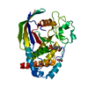 2cjzC  2gjtC  2h4vC  2i75C  2jjdC  2nlkC  2nz6C  2oc3C  2ooqC  2p6xC  2pa5C  2qepC  2shpS S: Starting model for refinement C: citing same article ( |
|---|---|
| Similar structure data |


-Links
 PDBj
PDBj


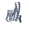
- Assembly
| Deposited unit | 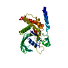
| ||||||||
|---|---|---|---|---|---|---|---|---|---|
| 1 |
| ||||||||
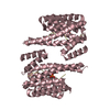
| Unit cell |
|
-Components
| #1: Protein | Mass: 36879.723 Da / Num. of mol.: 1 / Fragment: 'Residues 237-529 (Isoform 2)' Source method: isolated from a genetically manipulated source Source: (gene. exp.) Homo sapiens (human) / Gene: PTPN11, PTP2C, SHPTP2 / Plasmid: pNIC28-Bsa4 / Production host:  |
|---|---|
| #2: Chemical | ChemComp-MLT /   Mass: 134.087 Da / Num. of mol.: 1 / Source method: obtained synthetically / Formula: C4H6O5 Mass: 134.087 Da / Num. of mol.: 1 / Source method: obtained synthetically / Formula: C4H6O5 |
| #3: Water | ChemComp-HOH /  Mass: 18.015 Da / Num. of mol.: 241 / Source method: isolated from a natural source / Formula: H2O Mass: 18.015 Da / Num. of mol.: 241 / Source method: isolated from a natural source / Formula: H2O |
-Experimental details
-Experiment
| Experiment | Method: X-RAY DIFFRACTION / Number of used crystals: 1 |
|---|
- Sample preparation
| Crystal | Density Matthews: 2.19 Å3/Da / Density % sol: 43.79 % |
|---|---|
| Crystal grow | Temperature: 293 K / Method: vapor diffusion, sitting drop / pH: 7.5 Details: 20% PEG 3350, 0.15 M Na Malate, pH 7.5, VAPOR DIFFUSION, SITTING DROP, temperature 293K |
-Data collection
| Diffraction | Mean temperature: 100 K |
|---|---|
| Diffraction source | Source: SYNCHROTRON / Site: SLS  / Beamline: X10SA / Wavelength: 1.00721 Å / Beamline: X10SA / Wavelength: 1.00721 Å |
| Detector | Type: MARMOSAIC 225 mm CCD / Detector: CCD / Date: Jul 15, 2007 |
| Radiation | Protocol: SINGLE WAVELENGTH / Monochromatic (M) / Laue (L): M / Scattering type: x-ray |
| Radiation wavelength | Wavelength: 1.00721 Å / Relative weight: 1 |
| Reflection | Resolution: 1.6→34.12 Å / Num. all: 43184 / Num. obs: 43184 / % possible obs: 99.9 % / Observed criterion σ(F): 0 / Observed criterion σ(I): 0 / Redundancy: 4.1 % / Biso Wilson estimate: 19.1 Å2 / Rmerge(I) obs: 0.092 / Rsym value: 0.092 / Net I/σ(I): 13.3 |
| Reflection shell | Resolution: 1.6→1.69 Å / Redundancy: 4 % / Rmerge(I) obs: 1.01 / Mean I/σ(I) obs: 2 / Num. unique all: 25196 / Rsym value: 1.01 / % possible all: 100 |
- Processing
| Software |
| |||||||||||||||||||||||||||||||||||||||||||||||||||||||||||||||||||||||||||||||||||||||||||||||||||||||||||||||||||||||||||||
|---|---|---|---|---|---|---|---|---|---|---|---|---|---|---|---|---|---|---|---|---|---|---|---|---|---|---|---|---|---|---|---|---|---|---|---|---|---|---|---|---|---|---|---|---|---|---|---|---|---|---|---|---|---|---|---|---|---|---|---|---|---|---|---|---|---|---|---|---|---|---|---|---|---|---|---|---|---|---|---|---|---|---|---|---|---|---|---|---|---|---|---|---|---|---|---|---|---|---|---|---|---|---|---|---|---|---|---|---|---|---|---|---|---|---|---|---|---|---|---|---|---|---|---|---|---|---|
| Refinement | Method to determine structure: MOLECULAR REPLACEMENT Starting model: PDB entry 2SHP Resolution: 1.6→34.12 Å / Cor.coef. Fo:Fc: 0.967 / Cor.coef. Fo:Fc free: 0.95 / SU B: 3.422 / SU ML: 0.058 / TLS residual ADP flag: LIKELY RESIDUAL / Cross valid method: THROUGHOUT / σ(F): 0 / ESU R: 0.079 / ESU R Free: 0.085 / Stereochemistry target values: MAXIMUM LIKELIHOOD / Details: HYDROGENS HAVE BEEN ADDED IN THE RIDING POSITIONS
| |||||||||||||||||||||||||||||||||||||||||||||||||||||||||||||||||||||||||||||||||||||||||||||||||||||||||||||||||||||||||||||
| Solvent computation | Ion probe radii: 0.8 Å / Shrinkage radii: 0.8 Å / VDW probe radii: 1.2 Å / Solvent model: MASK | |||||||||||||||||||||||||||||||||||||||||||||||||||||||||||||||||||||||||||||||||||||||||||||||||||||||||||||||||||||||||||||
| Displacement parameters | Biso mean: 16.903 Å2
| |||||||||||||||||||||||||||||||||||||||||||||||||||||||||||||||||||||||||||||||||||||||||||||||||||||||||||||||||||||||||||||
| Refinement step | Cycle: LAST / Resolution: 1.6→34.12 Å
| |||||||||||||||||||||||||||||||||||||||||||||||||||||||||||||||||||||||||||||||||||||||||||||||||||||||||||||||||||||||||||||
| Refine LS restraints |
| |||||||||||||||||||||||||||||||||||||||||||||||||||||||||||||||||||||||||||||||||||||||||||||||||||||||||||||||||||||||||||||
| LS refinement shell | Resolution: 1.6→1.642 Å / Total num. of bins used: 20
| |||||||||||||||||||||||||||||||||||||||||||||||||||||||||||||||||||||||||||||||||||||||||||||||||||||||||||||||||||||||||||||
| Refinement TLS params. | Method: refined / Origin x: 17.0831 Å / Origin y: 10.0483 Å / Origin z: 63.979 Å
|
