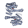
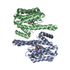
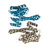
- PDB-3ual: Crystal Structure of 14-3-3 epsilon with Mlf1 peptide -
+
Open data
ID or keywords:
Loading...
-
Basic information
Entry
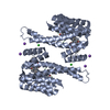
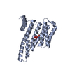
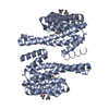
Database: PDB / ID: 3ual
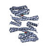
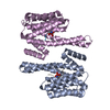
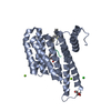
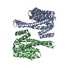
Title
Crystal Structure of 14-3-3 epsilon with Mlf1 peptide
Components
14-3-3 protein epsilon
Myeloid leukemia factor 1
Keywords
SIGNALING PROTEIN/PROTEIN BINDING / Adapter protein / ALL HELICAL / PHOSPHOPETIDE / SIGNALING PROTEIN-PROTEIN BINDING complex
Function / homology
Function and homology information
regulation of heart rate by hormone / positive regulation of hippo signaling / membrane repolarization during cardiac muscle cell action potential / regulation of cell cycle G1/S phase transition / negative regulation of toll-like receptor signaling pathway / regulation of membrane repolarization / protein localization to endoplasmic reticulum / NADE modulates death signalling / myeloid progenitor cell differentiation / RAB GEFs exchange GTP for GDP on RABs ...regulation of heart rate by hormone / positive regulation of hippo signaling / membrane repolarization during cardiac muscle cell action potential / regulation of cell cycle G1/S phase transition / negative regulation of toll-like receptor signaling pathway / regulation of membrane repolarization / protein localization to endoplasmic reticulum / NADE modulates death signalling / myeloid progenitor cell differentiation / RAB GEFs exchange GTP for GDP on RABs / Signaling by Hippo / regulation of potassium ion transmembrane transport / protein phosphatase inhibitor activity / Deregulated CDK5 triggers multiple neurodegenerative pathways in Alzheimer's disease models / negative regulation of calcium ion export across plasma membrane / cytoplasmic pattern recognition receptor signaling pathway / intracellular potassium ion homeostasis / regulation of heart rate by cardiac conduction / Regulation of HSF1-mediated heat shock response / phosphoserine residue binding / Activation of BAD and translocation to mitochondria / potassium channel regulator activity / HSF1 activation / SARS-CoV-2 targets host intracellular signalling and regulatory pathways / protein localization to nucleus / regulation of cytosolic calcium ion concentration / SARS-CoV-1 targets host intracellular signalling and regulatory pathways / calcium channel inhibitor activity / RHO GTPases activate PKNs / protein targeting / Chk1/Chk2(Cds1) mediated inactivation of Cyclin B:Cdk1 complex / Loss of Nlp from mitotic centrosomes / Loss of proteins required for interphase microtubule organization from the centrosome / Transcriptional and post-translational regulation of MITF-M expression and activity / Recruitment of mitotic centrosome proteins and complexes / Recruitment of NuMA to mitotic centrosomes / Anchoring of the basal body to the plasma membrane / signaling adaptor activity / substantia nigra development / regulation of mitotic cell cycle / AURKA Activation by TPX2 / positive regulation of protein export from nucleus / regulation of signal transduction by p53 class mediator / TP53 Regulates Metabolic Genes / Translocation of SLC2A4 (GLUT4) to the plasma membrane / hippocampus development / calcium channel regulator activity / protein sequestering activity / phosphoprotein binding / cerebral cortex development / mitochondrial membrane / neuron migration / histone deacetylase binding / melanosome / MHC class II protein complex binding / intracellular protein localization / Regulation of PLK1 Activity at G2/M Transition / MAPK cascade / cellular response to heat / scaffold protein binding / protein phosphatase binding / transmembrane transporter binding / intracellular signal transduction / cilium / ciliary basal body / cadherin binding / protein heterodimerization activity / protein domain specific binding / focal adhesion / ubiquitin protein ligase binding / regulation of DNA-templated transcription / DNA-templated transcription / perinuclear region of cytoplasm / enzyme binding / signal transduction / endoplasmic reticulum / DNA binding / RNA binding / extracellular exosome / membrane / identical protein binding / nucleus / cytoplasm / cytosol Similarity search - Function
Resolution: 1.8→35.27 Å / Cor.coef. Fo:Fc: 0.964 / Cor.coef. Fo:Fc free: 0.937 / SU B: 4.996 / SU ML: 0.077 / Cross valid method: THROUGHOUT / σ(F): 0 / ESU R Free: 0.125 / Stereochemistry target values: MAXIMUM LIKELIHOOD / Details: HYDROGENS HAVE BEEN ADDED IN THE RIDING POSITIONS
Rfactor
Num. reflection
% reflection
Selection details
Rfree
0.2224
1215
5 %
RANDOM
Rwork
0.1719
-
-
-
all
0.17442
24508
-
-
obs
0.17442
23072
100 %
-
Solvent computation
Ion probe radii: 0.8 Å / Shrinkage radii: 0.8 Å / VDW probe radii: 1.2 Å / Solvent model: MASK
Displacement parameters
Biso mean: 32.07 Å2
Baniso -1
Baniso -2
Baniso -3
1-
-1.24 Å2
0 Å2
0 Å2
2-
-
-0.75 Å2
0 Å2
3-
-
-
1.99 Å2
Refinement step
Cycle: LAST / Resolution: 1.8→35.27 Å
Protein
Nucleic acid
Ligand
Solvent
Total
Num. atoms
1912
0
15
159
2086
Refine LS restraints
Refine-ID
Type
Dev ideal
Dev ideal target
Number
X-RAY DIFFRACTION
r_bond_refined_d
0.023
0.022
2059
X-RAY DIFFRACTION
r_bond_other_d
X-RAY DIFFRACTION
r_angle_refined_deg
1.909
1.988
2801
X-RAY DIFFRACTION
r_angle_other_deg
X-RAY DIFFRACTION
r_dihedral_angle_1_deg
5.282
5
266
X-RAY DIFFRACTION
r_dihedral_angle_2_deg
36.329
24.423
104
X-RAY DIFFRACTION
r_dihedral_angle_3_deg
15.051
15.038
395
X-RAY DIFFRACTION
r_dihedral_angle_4_deg
17.583
15
17
X-RAY DIFFRACTION
r_chiral_restr
0.141
0.2
310
X-RAY DIFFRACTION
r_gen_planes_refined
0.01
0.021
1560
X-RAY DIFFRACTION
r_gen_planes_other
X-RAY DIFFRACTION
r_nbd_refined
X-RAY DIFFRACTION
r_nbd_other
X-RAY DIFFRACTION
r_nbtor_refined
X-RAY DIFFRACTION
r_nbtor_other
X-RAY DIFFRACTION
r_xyhbond_nbd_refined
X-RAY DIFFRACTION
r_xyhbond_nbd_other
X-RAY DIFFRACTION
r_metal_ion_refined
X-RAY DIFFRACTION
r_metal_ion_other
X-RAY DIFFRACTION
r_symmetry_vdw_refined
X-RAY DIFFRACTION
r_symmetry_vdw_other
X-RAY DIFFRACTION
r_symmetry_hbond_refined
X-RAY DIFFRACTION
r_symmetry_hbond_other
X-RAY DIFFRACTION
r_symmetry_metal_ion_refined
X-RAY DIFFRACTION
r_symmetry_metal_ion_other
X-RAY DIFFRACTION
r_mcbond_it
1.233
1.5
1251
X-RAY DIFFRACTION
r_mcbond_other
X-RAY DIFFRACTION
r_mcangle_it
2.003
2
2017
X-RAY DIFFRACTION
r_scbond_it
3.48
3
808
X-RAY DIFFRACTION
r_scangle_it
5.484
4.5
770
X-RAY DIFFRACTION
r_rigid_bond_restr
X-RAY DIFFRACTION
r_sphericity_free
X-RAY DIFFRACTION
r_sphericity_bonded
LS refinement shell
Resolution: 1.8→1.847 Å / Total num. of bins used: 20
Rfactor
Num. reflection
% reflection
Rfree
0.259
88
-
Rwork
0.21
1671
-
obs
-
-
100 %
Refinement TLS params.
Method: refined / Origin x: -18.8512 Å / Origin y: 5.6095 Å / Origin z: 20.9416 Å
11
12
13
21
22
23
31
32
33
T
0.0446 Å2
0.0275 Å2
0.0005 Å2
-
0.0213 Å2
-0.012 Å2
-
-
0.0728 Å2
L
2.201 °2
0.5629 °2
0.3282 °2
-
1.2322 °2
0.2857 °2
-
-
1.4513 °2
S
-0.0666 Å °
-0.022 Å °
0.0977 Å °
0.0416 Å °
-0.0204 Å °
0.1128 Å °
-0.0324 Å °
-0.0162 Å °
0.087 Å °
+
About Yorodumi
-
News
-
Feb 9, 2022. New format data for meta-information of EMDB entries
New format data for meta-information of EMDB entries
Version 3 of the EMDB header file is now the official format.
The previous official version 1.9 will be removed from the archive.
In the structure databanks used in Yorodumi, some data are registered as the other names, "COVID-19 virus" and "2019-nCoV". Here are the details of the virus and the list of structure data.
Jan 31, 2019. EMDB accession codes are about to change! (news from PDBe EMDB page)
EMDB accession codes are about to change! (news from PDBe EMDB page)
The allocation of 4 digits for EMDB accession codes will soon come to an end. Whilst these codes will remain in use, new EMDB accession codes will include an additional digit and will expand incrementally as the available range of codes is exhausted. The current 4-digit format prefixed with “EMD-” (i.e. EMD-XXXX) will advance to a 5-digit format (i.e. EMD-XXXXX), and so on. It is currently estimated that the 4-digit codes will be depleted around Spring 2019, at which point the 5-digit format will come into force.
The EM Navigator/Yorodumi systems omit the EMD- prefix.
Related info.:Q: What is EMD? / ID/Accession-code notation in Yorodumi/EM Navigator
Yorodumi is a browser for structure data from EMDB, PDB, SASBDB, etc.
This page is also the successor to EM Navigator detail page, and also detail information page/front-end page for Omokage search.
The word "yorodu" (or yorozu) is an old Japanese word meaning "ten thousand". "mi" (miru) is to see.
Related info.:EMDB / PDB / SASBDB / Comparison of 3 databanks / Yorodumi Search / Aug 31, 2016. New EM Navigator & Yorodumi / Yorodumi Papers / Jmol/JSmol / Function and homology information / Changes in new EM Navigator and Yorodumi
 Movie
Movie Controller


 Open data
 Basic information
 Components
 Keywords
 Function and homology information
 Homo sapiens (human)
 X-RAY DIFFRACTION /
  Authors
 Citation
 Structure visualization
 Downloads & links
 Other downloads
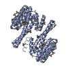


 PDBj
PDBj


 Assembly
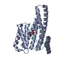

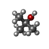
 Mass: 74.122 Da / Num. of mol.: 3 / Source method: obtained synthetically / Formula: C4H10O
 Mass: 18.015 Da / Num. of mol.: 159 / Source method: isolated from a natural source / Formula: H2O
 Sample preparation
 / Beamline: X10SA / Wavelength: 0.97906 Å
 Processing