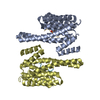
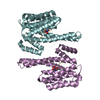
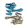
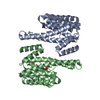
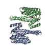
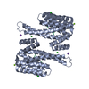
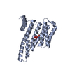
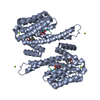
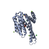
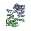
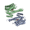
- PDB-6gkg: Structure of 14-3-3 gamma in complex with caspase-2 14-3-3 bindin... -
+
Open data
ID or keywords:
Loading...
-
Basic information
Entry
Database: PDB / ID: 6gkg
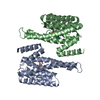
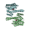
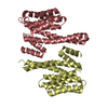
Title
Structure of 14-3-3 gamma in complex with caspase-2 14-3-3 binding motif Ser164
Components
14-3-3 protein gamma
Caspase-2
Keywords
SIGNALING PROTEIN / complex / phosphorylation / 14-3-3 protein / caspase-2
Function / homology
Function and homology information
caspase-2 / endopeptidase complex / positive regulation of cell-cell adhesion / phosphorylation-dependent protein binding / NADE modulates death signalling / luteolysis / neural retina development / positive regulation of T cell mediated immune response to tumor cell / regulation of neuron differentiation / execution phase of apoptosis ...caspase-2 / endopeptidase complex / positive regulation of cell-cell adhesion / phosphorylation-dependent protein binding / NADE modulates death signalling / luteolysis / neural retina development / positive regulation of T cell mediated immune response to tumor cell / regulation of neuron differentiation / execution phase of apoptosis / TP53 Regulates Transcription of Caspase Activators and Caspases / protein kinase C inhibitor activity / Regulation of localization of FOXO transcription factors / Activation of BAD and translocation to mitochondria / regulation of signal transduction / SARS-CoV-2 targets host intracellular signalling and regulatory pathways / ectopic germ cell programmed cell death / negative regulation of protein kinase activity / cellular response to glucose starvation / SARS-CoV-1 targets host intracellular signalling and regulatory pathways / RHO GTPases activate PKNs / protein targeting / Chk1/Chk2(Cds1) mediated inactivation of Cyclin B:Cdk1 complex / insulin-like growth factor receptor binding / extrinsic apoptotic signaling pathway in absence of ligand / negative regulation of TORC1 signaling / Loss of Nlp from mitotic centrosomes / Loss of proteins required for interphase microtubule organization from the centrosome / Transcriptional and post-translational regulation of MITF-M expression and activity / Recruitment of mitotic centrosome proteins and complexes / Recruitment of NuMA to mitotic centrosomes / Anchoring of the basal body to the plasma membrane / AURKA Activation by TPX2 / protein kinase C binding / TP53 Regulates Metabolic Genes / Translocation of SLC2A4 (GLUT4) to the plasma membrane / positive regulation of apoptotic signaling pathway / DNA damage response, signal transduction by p53 class mediator / protein sequestering activity / apoptotic signaling pathway / cellular response to mechanical stimulus / protein processing / NOD1/2 Signaling Pathway / receptor tyrosine kinase binding / regulation of synaptic plasticity / intrinsic apoptotic signaling pathway in response to DNA damage / positive regulation of T cell activation / cellular response to insulin stimulus / intracellular protein localization / Regulation of PLK1 Activity at G2/M Transition / positive regulation of neuron apoptotic process / regulation of protein localization / presynapse / positive regulation of apoptotic process / mitochondrial matrix / protein domain specific binding / cysteine-type endopeptidase activity / focal adhesion / apoptotic process / DNA damage response / negative regulation of apoptotic process / nucleolus / enzyme binding / signal transduction / RNA binding / extracellular exosome / membrane / identical protein binding / nucleus / cytoplasm / cytosol Similarity search - Function
Caspase-2 / Caspase recruitment domain / CARD domain / CARD caspase recruitment domain profile. / Caspase recruitment domain / Peptidase C14 family / 14-3-3 domain / Peptidase family C14A, His active site / Caspase family histidine active site. / Peptidase C14, caspase non-catalytic subunit p10 ...Caspase-2 / Caspase recruitment domain / CARD domain / CARD caspase recruitment domain profile. / Caspase recruitment domain / Peptidase C14 family / 14-3-3 domain / Peptidase family C14A, His active site / Caspase family histidine active site. / Peptidase C14, caspase non-catalytic subunit p10 / Peptidase family C14A, cysteine active site / Caspase family cysteine active site. / Caspase family p10 domain profile. / Peptidase C14A, caspase catalytic domain / Caspase, interleukin-1 beta converting enzyme (ICE) homologues / Delta-Endotoxin; domain 1 / Peptidase C14, p20 domain / Caspase family p20 domain profile. / : / Caspase domain / Caspase-like domain superfamily / 14-3-3 proteins signature 2. / 14-3-3 protein, conserved site / 14-3-3 proteins signature 1. / 14-3-3 protein / 14-3-3 homologues / 14-3-3 domain / 14-3-3 domain superfamily / 14-3-3 protein / Death-like domain superfamily / Up-down Bundle / Mainly Alpha Similarity search - Domain/homology
In the structure databanks used in Yorodumi, some data are registered as the other names, "COVID-19 virus" and "2019-nCoV". Here are the details of the virus and the list of structure data.
Jan 31, 2019. EMDB accession codes are about to change! (news from PDBe EMDB page)
EMDB accession codes are about to change! (news from PDBe EMDB page)
The allocation of 4 digits for EMDB accession codes will soon come to an end. Whilst these codes will remain in use, new EMDB accession codes will include an additional digit and will expand incrementally as the available range of codes is exhausted. The current 4-digit format prefixed with “EMD-” (i.e. EMD-XXXX) will advance to a 5-digit format (i.e. EMD-XXXXX), and so on. It is currently estimated that the 4-digit codes will be depleted around Spring 2019, at which point the 5-digit format will come into force.
The EM Navigator/Yorodumi systems omit the EMD- prefix.
Related info.:Q: What is EMD? / ID/Accession-code notation in Yorodumi/EM Navigator
Yorodumi is a browser for structure data from EMDB, PDB, SASBDB, etc.
This page is also the successor to EM Navigator detail page, and also detail information page/front-end page for Omokage search.
The word "yorodu" (or yorozu) is an old Japanese word meaning "ten thousand". "mi" (miru) is to see.
Related info.:EMDB / PDB / SASBDB / Comparison of 3 databanks / Yorodumi Search / Aug 31, 2016. New EM Navigator & Yorodumi / Yorodumi Papers / Jmol/JSmol / Function and homology information / Changes in new EM Navigator and Yorodumi
 Movie
Movie Controller

 Yorodumi
 Open data
 Basic information
 Components
 Keywords
 Function and homology information
 Homo sapiens (human)
 X-RAY DIFFRACTION /
  Authors
 Czech Republic, 1items
  Citation
 Structure visualization
 Downloads & links
 Other downloads
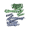


 PDBj
PDBj


 Assembly
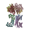


 Sample preparation
 / Beamline: 14.2 / Wavelength: 0.9184 Å
 Processing