[English] 日本語
Yorodumi
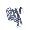
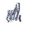
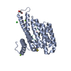
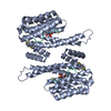
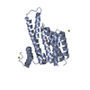
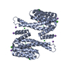
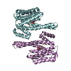
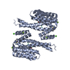
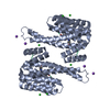
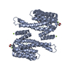
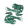
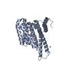
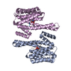
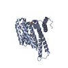
- PDB-5lu2: Human 14-3-3 sigma complexed with long HSPB6 phosphopeptide -

+
Open data


ID or keywords:

Loading...

-
Basic information

Entry
Database: PDB / ID: 5lu2
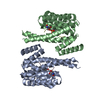
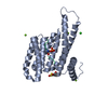
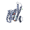
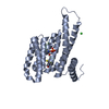
TitleHuman 14-3-3 sigma complexed with long HSPB6 phosphopeptide
Components
  • 14-3-3 protein sigma
  • Heat shock protein beta-6Heat shock response
KeywordsSIGNALING PROTEIN / protein-peptide complex / intrinsically disordered protein region(s)
Function / homology
Function and homology information


structural constituent of eye lens / regulation of epidermal cell division / protein kinase C inhibitor activity / positive regulation of epidermal cell differentiation / keratinocyte development / keratinization / Regulation of localization of FOXO transcription factors / keratinocyte proliferation / phosphoserine residue binding / Activation of BAD and translocation to mitochondria ...structural constituent of eye lens / regulation of epidermal cell division / protein kinase C inhibitor activity / positive regulation of epidermal cell differentiation / keratinocyte development / keratinization / Regulation of localization of FOXO transcription factors / keratinocyte proliferation / phosphoserine residue binding / Activation of BAD and translocation to mitochondria / negative regulation of keratinocyte proliferation / establishment of skin barrier / SARS-CoV-2 targets host intracellular signalling and regulatory pathways / Chk1/Chk2(Cds1) mediated inactivation of Cyclin B:Cdk1 complex / chaperone-mediated protein folding / protein folding chaperone / protein kinase A signaling / negative regulation of stem cell proliferation / SARS-CoV-1 targets host intracellular signalling and regulatory pathways / RHO GTPases activate PKNs / protein export from nucleus / negative regulation of innate immune response / protein sequestering activity / TP53 Regulates Transcription of Genes Involved in G2 Cell Cycle Arrest / release of cytochrome c from mitochondria / positive regulation of protein export from nucleus / stem cell proliferation / Translocation of SLC2A4 (GLUT4) to the plasma membrane / TP53 Regulates Metabolic Genes / negative regulation of protein kinase activity / negative regulation of cysteine-type endopeptidase activity involved in apoptotic process / positive regulation of angiogenesis / intrinsic apoptotic signaling pathway in response to DNA damage / unfolded protein binding / response to heat / protein-folding chaperone binding / protein refolding / positive regulation of cell growth / regulation of cell cycle / nuclear speck / cadherin binding / negative regulation of apoptotic process / protein kinase binding / negative regulation of transcription by RNA polymerase II / signal transduction / protein homodimerization activity / mitochondrion / extracellular space / extracellular exosome / extracellular region / identical protein binding / nucleus / cytosol / cytoplasm
Similarity search - Function
Alpha-crystallin, N-terminal / Alpha crystallin A chain, N terminal / Alpha crystallin/Small heat shock protein, animal type / Hsp20/alpha crystallin family / Small heat shock protein (sHSP) domain profile. / Alpha crystallin/Hsp20 domain / HSP20-like chaperone / 14-3-3 domain / Delta-Endotoxin; domain 1 / 14-3-3 protein sigma ...Alpha-crystallin, N-terminal / Alpha crystallin A chain, N terminal / Alpha crystallin/Small heat shock protein, animal type / Hsp20/alpha crystallin family / Small heat shock protein (sHSP) domain profile. / Alpha crystallin/Hsp20 domain / HSP20-like chaperone / 14-3-3 domain / Delta-Endotoxin; domain 1 / 14-3-3 protein sigma / 14-3-3 proteins signature 2. / 14-3-3 protein, conserved site / 14-3-3 proteins signature 1. / 14-3-3 protein / 14-3-3 homologues / 14-3-3 domain / 14-3-3 domain superfamily / 14-3-3 protein / Up-down Bundle / Mainly Alpha
Similarity search - Domain/homology
Heat shock protein beta-6 / 14-3-3 protein sigma
Similarity search - Component
Biological speciesHomo sapiens (human)
MethodX-RAY DIFFRACTION / SYNCHROTRON / MOLECULAR REPLACEMENT / Resolution: 2.5 Å
AuthorsSluchanko, N.N. / Beelen, S. / Kulikova, A.A. / Weeks, S.D. / Antson, A.A. / Gusev, N.B. / Strelkov, S.V.
CitationJournal: Structure / Year: 2017
Title: Structural Basis for the Interaction of a Human Small Heat Shock Protein with the 14-3-3 Universal Signaling Regulator.
Authors: Sluchanko, N.N. / Beelen, S. / Kulikova, A.A. / Weeks, S.D. / Antson, A.A. / Gusev, N.B. / Strelkov, S.V.
History
DepositionSep 7, 2016Deposition site: PDBE / Processing site: PDBE
Revision 1.0Feb 1, 2017Provider: repository / Type: Initial release
Revision 1.1Feb 15, 2017Group: Database references
Revision 1.2Jan 17, 2024Group: Data collection / Database references / Refinement description
Category: chem_comp_atom / chem_comp_bond ...chem_comp_atom / chem_comp_bond / database_2 / pdbx_initial_refinement_model
Item: _database_2.pdbx_DOI / _database_2.pdbx_database_accession

-
Structure visualization

Structure viewerMolecule:
MolmilJmol/JSmol

Downloads & links

-
Assembly

Deposited unit
A: 14-3-3 protein sigma
B: 14-3-3 protein sigma
C: Heat shock protein beta-6
D: Heat shock protein beta-6


Theoretical massNumber of molelcules
Total (without water)55,3744
Polymers55,3744
Non-polymers00
Water2,288127
1


  • Idetical with deposited unit
  • defined by author&software
TypeNameSymmetry operationNumber
identity operation1_555x,y,z1
Buried area4510 Å2
ΔGint-27 kcal/mol
Surface area22180 Å2
MethodPISA
Unit cell
Length a, b, c (Å)90.060, 78.980, 75.360
Angle α, β, γ (deg.)90.000, 102.470, 90.000
Int Tables number5
Space group name H-MC121

-
Components

#1: Protein 14-3-3 protein sigma / Sigma WT adaptor / Epithelial cell marker protein 1 / Stratifin


Mass: 26139.461 Da / Num. of mol.: 2 / Fragment: UNP residues 1-231
Source method: isolated from a genetically manipulated source
Source: (gene. exp.) Homo sapiens (human) / Gene: SFN, HME1 / Production host: Escherichia coli (E. coli) / References: UniProt: P31947
#2: Protein/peptide Heat shock protein beta-6 / Heat shock response / HspB6 / Heat shock 20 kDa-like protein p20


Mass: 1547.760 Da / Num. of mol.: 2 / Fragment: UNP residues 11-23 / Source method: obtained synthetically / Source: (synth.) Homo sapiens (human) / References: UniProt: O14558
#3: Water ChemComp-HOH / water / Water


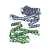
Mass: 18.015 Da / Num. of mol.: 127 / Source method: isolated from a natural source / Formula: H2O

-
Experimental details

-
Experiment

ExperimentMethod: X-RAY DIFFRACTION / Number of used crystals: 1

-
Sample preparation

CrystalDensity Matthews: 2.36 Å3/Da / Density % sol: 47.95 %
Crystal growTemperature: 277 K / Method: vapor diffusion, sitting drop / pH: 7.5 / Details: 0.1 M HEPES (pH 7.5), 0.2 M MgCl2 and 30% PEG 400

-
Data collection

DiffractionMean temperature: 100 K
Diffraction sourceSource: SYNCHROTRON / Site: SOLEIL / Beamline: PROXIMA 2 / Wavelength: 0.9801 Å
DetectorType: ADSC QUANTUM 315 / Detector: CCD / Date: Jun 12, 2015
RadiationProtocol: SINGLE WAVELENGTH / Monochromatic (M) / Laue (L): M / Scattering type: x-ray
Radiation wavelengthWavelength: 0.9801 Å / Relative weight: 1
ReflectionResolution: 2.5→44 Å / Num. obs: 17880 / % possible obs: 99.6 % / Observed criterion σ(I): -3 / Redundancy: 3.7 % / Biso Wilson estimate: 80.39 Å2 / CC1/2: 0.994 / Rmerge(I) obs: 0.15 / Net I/σ(I): 7.11
Reflection shell
Resolution (Å)Highest resolution (Å)Rmerge(I) obsMean I/σ(I) obsCC1/2Diffraction-ID% possible all
2.5-2.561.8170.880.261100
2.56-2.641.5371.050.363199.8
2.64-2.711.2621.340.445199.9
2.71-2.80.971.720.659199.8
2.8-2.890.8152.080.735199.6
2.89-2.990.7312.530.761199.7
2.99-3.10.6053.170.789199.8
3.1-3.230.4924.30.832199.3
3.23-3.370.3566.050.89199.9
3.37-3.540.2387.720.954199.7
3.54-3.730.1749.480.974199.6
3.73-3.950.12511.850.986199.7
3.95-4.230.09314.060.989199.3
4.23-4.560.07915.560.991198.8
4.56-50.07815.710.992198.6
5-5.590.08314.060.99198.9
5.59-6.460.07315.620.99199.5
6.46-7.910.05419.160.995199.4
7.91-11.180.03622.090.9971100
11.180.04422.410.996195.9

-
Processing

Software
NameVersionClassification
XSCALEdata scaling
BUSTER-TNT2.10.3refinement
PDB_EXTRACT3.2data extraction
XDSdata reduction
MOLREPphasing
RefinementMethod to determine structure: MOLECULAR REPLACEMENT
Starting model: 3IQJ
Resolution: 2.5→43.97 Å / Cor.coef. Fo:Fc: 0.94 / Cor.coef. Fo:Fc free: 0.913 / Rfactor Rfree error: 0 / SU R Cruickshank DPI: 0.67 / Cross valid method: THROUGHOUT / σ(F): 0 / SU R Blow DPI: 0.653 / SU Rfree Blow DPI: 0.285 / SU Rfree Cruickshank DPI: 0.291
RfactorNum. reflection% reflectionSelection details
Rfree0.25 1040 5.82 %RANDOM
Rwork0.209 ---
obs0.211 17876 99.6 %-
Displacement parametersBiso max: 168.74 Å2 / Biso mean: 74.57 Å2 / Biso min: 36.7 Å2
Baniso -1Baniso -2Baniso -3
1-5.1679 Å20 Å20.0114 Å2
2--3.1726 Å20 Å2
3----8.3405 Å2
Refine analyzeLuzzati coordinate error obs: 0.37 Å
Refinement stepCycle: final / Resolution: 2.5→43.97 Å
ProteinNucleic acidLigandSolventTotal
Num. atoms3753 0 0 127 3880
Biso mean---67.16 -
Num. residues----473
Refine LS restraints
Refine-IDTypeNumberRestraint functionWeightDev ideal
X-RAY DIFFRACTIONt_dihedral_angle_d1396SINUSOIDAL2
X-RAY DIFFRACTIONt_trig_c_planes115HARMONIC2
X-RAY DIFFRACTIONt_gen_planes538HARMONIC5
X-RAY DIFFRACTIONt_it3806HARMONIC20
X-RAY DIFFRACTIONt_nbd
X-RAY DIFFRACTIONt_improper_torsion
X-RAY DIFFRACTIONt_pseud_angle
X-RAY DIFFRACTIONt_chiral_improper_torsion490SEMIHARMONIC5
X-RAY DIFFRACTIONt_sum_occupancies
X-RAY DIFFRACTIONt_utility_distance
X-RAY DIFFRACTIONt_utility_angle
X-RAY DIFFRACTIONt_utility_torsion
X-RAY DIFFRACTIONt_ideal_dist_contact4422SEMIHARMONIC4
X-RAY DIFFRACTIONt_bond_d3806HARMONIC20.01
X-RAY DIFFRACTIONt_angle_deg5124HARMONIC21.19
X-RAY DIFFRACTIONt_omega_torsion2.66
X-RAY DIFFRACTIONt_other_torsion20.03
LS refinement shellResolution: 2.5→2.65 Å / Total num. of bins used: 9
RfactorNum. reflection% reflection
Rfree0.291 158 5.51 %
Rwork0.247 2712 -
all-2870 -
obs--99.9 %
Refinement TLS params.

Method: refined / Refine-ID: X-RAY DIFFRACTION

IDL112)L122)L132)L222)L232)L332)S11 (Å °)S12 (Å °)S13 (Å °)S21 (Å °)S22 (Å °)S23 (Å °)S31 (Å °)S32 (Å °)S33 (Å °)T112)T122)T132)T222)T232)T332)Origin x (Å)Origin y (Å)Origin z (Å)
12.25930.50060.24522.2302-0.25431.55910.18990.0626-0.3062-0.15560.0026-0.00660.331-0.0101-0.1925-0.2022-0.0048-0.0504-0.10240.0277-0.062830.2243-18.289720.1833
21.3007-0.19820.11382.8095-0.83711.23730.00750.05970.08020.0405-0.0069-0.0277-0.1758-0.0142-0.0006-0.18780.02730.07950.0267-0.0225-0.146831.325919.38120.4848
Refinement TLS group
IDRefine-IDRefine TLS-IDSelection detailsAuth asym-IDAuth seq-ID
1X-RAY DIFFRACTION1{ A|* D|* }A2 - 231
2X-RAY DIFFRACTION1{ A|* D|* }D13 - 20
3X-RAY DIFFRACTION2{ B|* C|* }B2 - 231
4X-RAY DIFFRACTION2{ B|* C|* }C13 - 20

+
About Yorodumi

-
News

-
Feb 9, 2022. New format data for meta-information of EMDB entries

New format data for meta-information of EMDB entries

  • Version 3 of the EMDB header file is now the official format.
  • The previous official version 1.9 will be removed from the archive.

Related info.:EMDB header

External links:wwPDB to switch to version 3 of the EMDB data model

-
Aug 12, 2020. Covid-19 info

Covid-19 info

URL: https://pdbj.org/emnavi/covid19.php

New page: Covid-19 featured information page in EM Navigator.

Related info.:Covid-19 info / Mar 5, 2020. Novel coronavirus structure data

+
Mar 5, 2020. Novel coronavirus structure data

Novel coronavirus structure data

Related info.:Yorodumi Speices / Aug 12, 2020. Covid-19 info

External links:COVID-19 featured content - PDBj / Molecule of the Month (242):Coronavirus Proteases

+
Jan 31, 2019. EMDB accession codes are about to change! (news from PDBe EMDB page)

EMDB accession codes are about to change! (news from PDBe EMDB page)

  • The allocation of 4 digits for EMDB accession codes will soon come to an end. Whilst these codes will remain in use, new EMDB accession codes will include an additional digit and will expand incrementally as the available range of codes is exhausted. The current 4-digit format prefixed with “EMD-” (i.e. EMD-XXXX) will advance to a 5-digit format (i.e. EMD-XXXXX), and so on. It is currently estimated that the 4-digit codes will be depleted around Spring 2019, at which point the 5-digit format will come into force.
  • The EM Navigator/Yorodumi systems omit the EMD- prefix.

Related info.:Q: What is EMD? / ID/Accession-code notation in Yorodumi/EM Navigator

External links:EMDB Accession Codes are Changing Soon! / Contact to PDBj

+
Jul 12, 2017. Major update of PDB

Major update of PDB

  • wwPDB released updated PDB data conforming to the new PDBx/mmCIF dictionary.
  • This is a major update changing the version number from 4 to 5, and with Remediation, in which all the entries are updated.
  • In this update, many items about electron microscopy experimental information are reorganized (e.g. em_software).
  • Now, EM Navigator and Yorodumi are based on the updated data.

External links:wwPDB Remediation / Enriched Model Files Conforming to OneDep Data Standards Now Available in the PDB FTP Archive

-
Yorodumi

Thousand views of thousand structures

  • Yorodumi is a browser for structure data from EMDB, PDB, SASBDB, etc.
  • This page is also the successor to EM Navigator detail page, and also detail information page/front-end page for Omokage search.
  • The word "yorodu" (or yorozu) is an old Japanese word meaning "ten thousand". "mi" (miru) is to see.

Related info.:EMDB / PDB / SASBDB / Comparison of 3 databanks / Yorodumi Search / Aug 31, 2016. New EM Navigator & Yorodumi / Yorodumi Papers / Jmol/JSmol / Function and homology information / Changes in new EM Navigator and Yorodumi

Read more