 Movie
Movie Controller

[English] 日本語
 Yorodumi
- PDB-5lu1: Human 14-3-3 sigma CLU3 mutant complexed with short HSPB6 phospho... -
+ Open data

- Basic information
| Entry | Database: PDB / ID: 5lu1 | ||||||
|---|---|---|---|---|---|---|---|
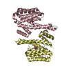
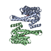
| Title | Human 14-3-3 sigma CLU3 mutant complexed with short HSPB6 phosphopeptide | ||||||
 Components Components |
| ||||||
 Keywords Keywords | SIGNALING PROTEIN / protein-peptide complex / intrinsically disordered protein region(s) | ||||||
| Function / homology |  Function and homology information Function and homology informationstructural constituent of eye lens / positive regulation of epidermal cell differentiation / keratinocyte development / regulation of epidermal cell division / protein kinase C inhibitor activity / keratinization / regulation of cell-cell adhesion / keratinocyte proliferation / establishment of skin barrier / Regulation of localization of FOXO transcription factors ...structural constituent of eye lens / positive regulation of epidermal cell differentiation / keratinocyte development / regulation of epidermal cell division / protein kinase C inhibitor activity / keratinization / regulation of cell-cell adhesion / keratinocyte proliferation / establishment of skin barrier / Regulation of localization of FOXO transcription factors / negative regulation of cardiac muscle cell apoptotic process / negative regulation of keratinocyte proliferation / phosphoserine residue binding / Activation of BAD and translocation to mitochondria / cAMP/PKA signal transduction / negative regulation of stem cell proliferation / negative regulation of protein localization to plasma membrane / SARS-CoV-2 targets host intracellular signalling and regulatory pathways / negative regulation of protein kinase activity / Chk1/Chk2(Cds1) mediated inactivation of Cyclin B:Cdk1 complex / SARS-CoV-1 targets host intracellular signalling and regulatory pathways / RHO GTPases activate PKNs / positive regulation of protein localization / stem cell proliferation / protein export from nucleus / protein folding chaperone / release of cytochrome c from mitochondria / TP53 Regulates Transcription of Genes Involved in G2 Cell Cycle Arrest / negative regulation of innate immune response / positive regulation of cell adhesion / positive regulation of protein export from nucleus / TP53 Regulates Metabolic Genes / Translocation of SLC2A4 (GLUT4) to the plasma membrane / protein sequestering activity / intrinsic apoptotic signaling pathway in response to DNA damage / positive regulation of angiogenesis / : / intracellular protein localization / regulation of protein localization / sperm midpiece / protein refolding / response to heat / protein-folding chaperone binding / positive regulation of cell growth / protein folding / regulation of cell cycle / nuclear speck / cadherin binding / protein kinase binding / negative regulation of apoptotic process / negative regulation of transcription by RNA polymerase II / signal transduction / protein homodimerization activity / mitochondrion / : / extracellular exosome / extracellular region / identical protein binding / nucleus / cytosol / cytoplasm Similarity search - Function | ||||||
| Biological species |  Homo sapiens (human) Homo sapiens (human) | ||||||
| Method |  X-RAY DIFFRACTION / SYNCHROTRON / MOLECULAR REPLACEMENT / Resolution: 2.4 Å X-RAY DIFFRACTION / SYNCHROTRON / MOLECULAR REPLACEMENT / Resolution: 2.4 Å | ||||||
 Authors Authors | Sluchanko, N.N. / Beelen, S. / Kulikova, A.A. / Weeks, S.D. / Antson, A.A. / Gusev, N.B. / Strelkov, S.V. | ||||||
 Citation Citation | Journal: Structure / Year: 2017 Title: Structural Basis for the Interaction of a Human Small Heat Shock Protein with the 14-3-3 Universal Signaling Regulator. Authors: Sluchanko, N.N. / Beelen, S. / Kulikova, A.A. / Weeks, S.D. / Antson, A.A. / Gusev, N.B. / Strelkov, S.V. | ||||||
| History |
|
- Structure visualization
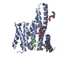
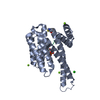
| Structure viewer | Molecule: MolmilJmol/JSmol |
|---|
- Downloads & links
-Download
| PDBx/mmCIF format | 5lu1.cif.gz | 390.5 KB | Display | PDBx/mmCIF format |
|---|---|---|---|---|
| PDB format | pdb5lu1.ent.gz | 323.4 KB | Display | PDB format |
| PDBx/mmJSON format | 5lu1.json.gz | Tree view | PDBx/mmJSON format | |
| Others |  Other downloads Other downloads |
-Validation report
| Arichive directory | https://data.pdbj.org/pub/pdb/validation_reports/lu/5lu1ftp://data.pdbj.org/pub/pdb/validation_reports/lu/5lu1 | HTTPS FTP |
|---|
-Related structure data
| Related structure data |  5ltwC 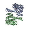 5lu2C  5lumC 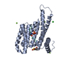 3iqjS S: Starting model for refinement C: citing same article ( |
|---|---|
| Similar structure data |


-Links
 PDBj
PDBj


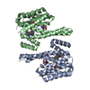
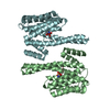
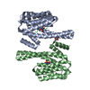
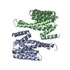
- Assembly
| Deposited unit | 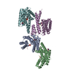
| ||||||||
|---|---|---|---|---|---|---|---|---|---|
| 1 | 
| ||||||||
| 2 | 
| ||||||||
| Unit cell |
|
-Components
| #1: Protein | Mass: 26257.600 Da / Num. of mol.: 4 / Fragment: UNP residues 1-231 / Mutation: CLU3 Source method: isolated from a genetically manipulated source Source: (gene. exp.) Homo sapiens (human) / Gene: SFN, HME1 / Production host:  #2: Protein/peptide | Mass: 949.003 Da / Num. of mol.: 4 / Fragment: UNP residues 13-20 / Source method: obtained synthetically / Source: (synth.) Homo sapiens (human) / References: UniProt: O14558#3: Chemical | ChemComp-TRS /   Mass: 122.143 Da / Num. of mol.: 4 / Source method: obtained synthetically / Formula: C4H12NO3 / Comment: pH buffer*YM Mass: 122.143 Da / Num. of mol.: 4 / Source method: obtained synthetically / Formula: C4H12NO3 / Comment: pH buffer*YM#4: Water | ChemComp-HOH / |  Mass: 18.015 Da / Num. of mol.: 361 / Source method: isolated from a natural source / Formula: H2O Mass: 18.015 Da / Num. of mol.: 361 / Source method: isolated from a natural source / Formula: H2OHas protein modification | Y | |
|---|
-Experimental details
-Experiment
| Experiment | Method: X-RAY DIFFRACTION / Number of used crystals: 1 |
|---|
- Sample preparation
| Crystal | Density Matthews: 3.26 Å3/Da / Density % sol: 62.3 % |
|---|---|
| Crystal grow | Temperature: 293 K / Method: vapor diffusion, sitting drop / pH: 6.5 Details: 0.1 M Bis-Tris propane (pH 6.5), 0.2 M NaNO3 and 20% PEG 3350 |
-Data collection
| Diffraction | Mean temperature: 100 K | |||||||||||||||||||||||||||||||||||||||||||||||||||||||||||||||||||||||||||||||||||||||||||||||||||||||||||||||||||||||||||||||||||||||||||||||||||
|---|---|---|---|---|---|---|---|---|---|---|---|---|---|---|---|---|---|---|---|---|---|---|---|---|---|---|---|---|---|---|---|---|---|---|---|---|---|---|---|---|---|---|---|---|---|---|---|---|---|---|---|---|---|---|---|---|---|---|---|---|---|---|---|---|---|---|---|---|---|---|---|---|---|---|---|---|---|---|---|---|---|---|---|---|---|---|---|---|---|---|---|---|---|---|---|---|---|---|---|---|---|---|---|---|---|---|---|---|---|---|---|---|---|---|---|---|---|---|---|---|---|---|---|---|---|---|---|---|---|---|---|---|---|---|---|---|---|---|---|---|---|---|---|---|---|---|---|---|
| Diffraction source | Source: SYNCHROTRON / Site: SOLEIL  / Beamline: PROXIMA 2 / Wavelength: 0.9801 Å / Beamline: PROXIMA 2 / Wavelength: 0.9801 Å | |||||||||||||||||||||||||||||||||||||||||||||||||||||||||||||||||||||||||||||||||||||||||||||||||||||||||||||||||||||||||||||||||||||||||||||||||||
| Detector | Type: ADSC QUANTUM 315 / Detector: CCD / Date: Jun 12, 2015 | |||||||||||||||||||||||||||||||||||||||||||||||||||||||||||||||||||||||||||||||||||||||||||||||||||||||||||||||||||||||||||||||||||||||||||||||||||
| Radiation | Protocol: SINGLE WAVELENGTH / Monochromatic (M) / Laue (L): M / Scattering type: x-ray | |||||||||||||||||||||||||||||||||||||||||||||||||||||||||||||||||||||||||||||||||||||||||||||||||||||||||||||||||||||||||||||||||||||||||||||||||||
| Radiation wavelength | Wavelength: 0.9801 Å / Relative weight: 1 | |||||||||||||||||||||||||||||||||||||||||||||||||||||||||||||||||||||||||||||||||||||||||||||||||||||||||||||||||||||||||||||||||||||||||||||||||||
| Reflection | Resolution: 2.4→48 Å / Num. obs: 54513 / % possible obs: 99.6 % / Observed criterion σ(I): -3 / Redundancy: 3.7 % / Biso Wilson estimate: 66.17 Å2 / CC1/2: 0.997 / Rmerge(I) obs: 0.099 / Net I/σ(I): 8.61 | |||||||||||||||||||||||||||||||||||||||||||||||||||||||||||||||||||||||||||||||||||||||||||||||||||||||||||||||||||||||||||||||||||||||||||||||||||
| Reflection shell |
|
- Processing
| Software |
| |||||||||||||||||||||||||||||||||||||||||||||||||||||||||||||||||||||||||||||||||||||||||||||||||||||||||||||||||||||||||||||
|---|---|---|---|---|---|---|---|---|---|---|---|---|---|---|---|---|---|---|---|---|---|---|---|---|---|---|---|---|---|---|---|---|---|---|---|---|---|---|---|---|---|---|---|---|---|---|---|---|---|---|---|---|---|---|---|---|---|---|---|---|---|---|---|---|---|---|---|---|---|---|---|---|---|---|---|---|---|---|---|---|---|---|---|---|---|---|---|---|---|---|---|---|---|---|---|---|---|---|---|---|---|---|---|---|---|---|---|---|---|---|---|---|---|---|---|---|---|---|---|---|---|---|---|---|---|---|
| Refinement | Method to determine structure: MOLECULAR REPLACEMENT Starting model: 3IQJ Resolution: 2.4→47.89 Å / Cor.coef. Fo:Fc: 0.949 / Cor.coef. Fo:Fc free: 0.938 / Rfactor Rfree error: 0 / SU R Cruickshank DPI: 0.257 / Cross valid method: THROUGHOUT / σ(F): 0 / SU R Blow DPI: 0.255 / SU Rfree Blow DPI: 0.192 / SU Rfree Cruickshank DPI: 0.195
| |||||||||||||||||||||||||||||||||||||||||||||||||||||||||||||||||||||||||||||||||||||||||||||||||||||||||||||||||||||||||||||
| Displacement parameters | Biso max: 201.42 Å2 / Biso mean: 72.11 Å2 / Biso min: 33.26 Å2
| |||||||||||||||||||||||||||||||||||||||||||||||||||||||||||||||||||||||||||||||||||||||||||||||||||||||||||||||||||||||||||||
| Refine analyze | Luzzati coordinate error obs: 0.31 Å | |||||||||||||||||||||||||||||||||||||||||||||||||||||||||||||||||||||||||||||||||||||||||||||||||||||||||||||||||||||||||||||
| Refinement step | Cycle: final / Resolution: 2.4→47.89 Å
| |||||||||||||||||||||||||||||||||||||||||||||||||||||||||||||||||||||||||||||||||||||||||||||||||||||||||||||||||||||||||||||
| Refine LS restraints |
| |||||||||||||||||||||||||||||||||||||||||||||||||||||||||||||||||||||||||||||||||||||||||||||||||||||||||||||||||||||||||||||
| LS refinement shell | Resolution: 2.4→2.46 Å / Total num. of bins used: 20
| |||||||||||||||||||||||||||||||||||||||||||||||||||||||||||||||||||||||||||||||||||||||||||||||||||||||||||||||||||||||||||||
| Refinement TLS params. | Method: refined / Refine-ID: X-RAY DIFFRACTION
| |||||||||||||||||||||||||||||||||||||||||||||||||||||||||||||||||||||||||||||||||||||||||||||||||||||||||||||||||||||||||||||
| Refinement TLS group |
|
