 Movie
Movie Controller


+ Open data

- Basic information
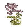
| Entry | Database: PDB / ID: 2c74 | ||||||
|---|---|---|---|---|---|---|---|
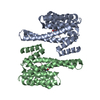
| Title | 14-3-3 Protein Eta (Human) Complexed to Peptide | ||||||
 Components Components |
| ||||||
 Keywords Keywords | SIGNALING PROTEIN/PEPTIDE / SIGNALING PROTEIN-PEPTIDE COMPLEX / 14-3-3 / PHOSPHOSERINE / PHOSPHORYLATION | ||||||
| Function / homology |  Function and homology information Function and homology informationglucocorticoid catabolic process / cerebellar granule cell to Purkinje cell synapse / nuclear receptor-mediated glucocorticoid signaling pathway / negative regulation of dendrite morphogenesis / nuclear glucocorticoid receptor binding / membrane depolarization during action potential / regulation of neuron differentiation / intercalated disc / sodium channel regulator activity / Activation of BAD and translocation to mitochondria ...glucocorticoid catabolic process / cerebellar granule cell to Purkinje cell synapse / nuclear receptor-mediated glucocorticoid signaling pathway / negative regulation of dendrite morphogenesis / nuclear glucocorticoid receptor binding / membrane depolarization during action potential / regulation of neuron differentiation / intercalated disc / sodium channel regulator activity / Activation of BAD and translocation to mitochondria / SARS-CoV-2 targets host intracellular signalling and regulatory pathways / SARS-CoV-1 targets host intracellular signalling and regulatory pathways / RHO GTPases activate PKNs / Chk1/Chk2(Cds1) mediated inactivation of Cyclin B:Cdk1 complex / regulation of sodium ion transport / insulin-like growth factor receptor binding / Transcriptional and post-translational regulation of MITF-M expression and activity / substantia nigra development / presynaptic modulation of chemical synaptic transmission / TP53 Regulates Metabolic Genes / Translocation of SLC2A4 (GLUT4) to the plasma membrane / intracellular protein transport / regulation of synaptic plasticity / intracellular protein localization / presynapse / actin binding / transmembrane transporter binding / protein heterodimerization activity / protein domain specific binding / positive regulation of DNA-templated transcription / enzyme binding / signal transduction / mitochondrion / extracellular exosome / identical protein binding / plasma membrane / cytoplasm / cytosol Similarity search - Function | ||||||
| Biological species |  HOMO SAPIENS (human) HOMO SAPIENS (human)synthetic construct (others) | ||||||
| Method |  X-RAY DIFFRACTION / SYNCHROTRON / MOLECULAR REPLACEMENT / Resolution: 2.7 Å X-RAY DIFFRACTION / SYNCHROTRON / MOLECULAR REPLACEMENT / Resolution: 2.7 Å | ||||||
 Authors Authors | Elkins, J.M. / Yang, X. / Smee, C.E.A. / Johansson, C. / Sundstrom, M. / Edwards, A. / Weigelt, J. / Arrowsmith, C. / Doyle, D.A. | ||||||
 Citation Citation | Journal: Proc. Natl. Acad. Sci. U.S.A. / Year: 2006 Title: Structural basis for protein-protein interactions in the 14-3-3 protein family. Authors: Yang, X. / Lee, W.H. / Sobott, F. / Papagrigoriou, E. / Robinson, C.V. / Grossmann, J.G. / Sundstrom, M. / Doyle, D.A. / Elkins, J.M. | ||||||
| History |
|
- Structure visualization
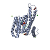
| Structure viewer | Molecule: MolmilJmol/JSmol |
|---|
- Downloads & links
-Download
| PDBx/mmCIF format | 2c74.cif.gz | 102.7 KB | Display | PDBx/mmCIF format |
|---|---|---|---|---|
| PDB format | pdb2c74.ent.gz | 78.3 KB | Display | PDB format |
| PDBx/mmJSON format | 2c74.json.gz | Tree view | PDBx/mmJSON format | |
| Others |  Other downloads Other downloads |
-Validation report
| Arichive directory | https://data.pdbj.org/pub/pdb/validation_reports/c7/2c74ftp://data.pdbj.org/pub/pdb/validation_reports/c7/2c74 | HTTPS FTP |
|---|
-Related structure data
| Related structure data | 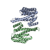 2bq0SC  2br9C 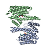 2btpC 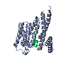 2c23C  2c63C C: citing same article ( S: Starting model for refinement |
|---|---|
| Similar structure data |


-Links
 PDBj
PDBj


- Assembly
| Deposited unit | 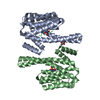
| ||||||||||||||||||||||||||||||||||||||||||||||||||
|---|---|---|---|---|---|---|---|---|---|---|---|---|---|---|---|---|---|---|---|---|---|---|---|---|---|---|---|---|---|---|---|---|---|---|---|---|---|---|---|---|---|---|---|---|---|---|---|---|---|---|---|
| 1 |
| ||||||||||||||||||||||||||||||||||||||||||||||||||
| Unit cell |
| ||||||||||||||||||||||||||||||||||||||||||||||||||
| Noncrystallographic symmetry (NCS) | NCS domain:
NCS domain segments:
NCS ensembles :
NCS oper: (Code: given Matrix: (-0.17433, -0.05026, 0.9834), Vector: Details | THE PROTEIN IS DIMERIC (CHAINS A,B) BUT SINCE EACH COMPONENT OF THIS DIMER IS IN COMPLEX WITH A PEPTIDE (CHAINSP,Q), THE ENTRY IS MARKED AS TETRAMERIC. | |
-Components
| #1: Protein | Mass: 28338.797 Da / Num. of mol.: 2 Source method: isolated from a genetically manipulated source Source: (gene. exp.) HOMO SAPIENS (human)Description: THE MAMMALIAN GENE COLLECTION, I.M.A.G.E. CONSORTIUM CLONEID 3543571 Plasmid: PNIC28-BSA4 / Production host:  #2: Protein/peptide | Mass: 952.975 Da / Num. of mol.: 2 / Source method: obtained synthetically / Details: PHOSPHOSERINE AT RESIDUE P 5, Q 5 / Source: (synth.) synthetic construct (others) #3: Chemical | ChemComp-CIT / |   Mass: 192.124 Da / Num. of mol.: 1 / Source method: obtained synthetically / Formula: C6H8O7 Mass: 192.124 Da / Num. of mol.: 1 / Source method: obtained synthetically / Formula: C6H8O7#4: Water | ChemComp-HOH / |  Mass: 18.015 Da / Num. of mol.: 3 / Source method: isolated from a natural source / Formula: H2O Mass: 18.015 Da / Num. of mol.: 3 / Source method: isolated from a natural source / Formula: H2OCompound details | INVOLVED IN THE REGULATION | Has protein modification | Y | |
|---|
-Experimental details
-Experiment
| Experiment | Method: X-RAY DIFFRACTION / Number of used crystals: 1 |
|---|
- Sample preparation
| Crystal | Density Matthews: 3.15 Å3/Da / Density % sol: 60.66 % |
|---|---|
| Crystal grow | pH: 5.6 / Details: 0.1M CITRATE PH 5.6, 20% ISOPROPANOL, 20% PEG4000 |
-Data collection
| Diffraction | Mean temperature: 100 K |
|---|---|
| Diffraction source | Source: SYNCHROTRON / Site: SLS  / Beamline: X10SA / Wavelength: 0.9789 / Beamline: X10SA / Wavelength: 0.9789 |
| Detector | Type: MARRESEARCH / Detector: CCD / Date: Oct 1, 2005 |
| Radiation | Protocol: SINGLE WAVELENGTH / Monochromatic (M) / Laue (L): M / Scattering type: x-ray |
| Radiation wavelength | Wavelength: 0.9789 Å / Relative weight: 1 |
| Reflection | Resolution: 2.7→38.9 Å / Num. obs: 18419 / % possible obs: 99.8 % / Observed criterion σ(I): 0 / Redundancy: 3.5 % / Rmerge(I) obs: 0.08 / Net I/σ(I): 12 |
| Reflection shell | Resolution: 2.7→2.85 Å / Redundancy: 3.6 % / Rmerge(I) obs: 0.48 / Mean I/σ(I) obs: 2.4 / % possible all: 99.9 |
- Processing
| Software |
| ||||||||||||||||||||||||||||||||||||||||||||||||||||||||||||||||||||||||||||||||||||||||||||||||||||||||||||||||||||||||||||||||||||||||||||||||||||||||||||||||||||||||||||||||||||||
|---|---|---|---|---|---|---|---|---|---|---|---|---|---|---|---|---|---|---|---|---|---|---|---|---|---|---|---|---|---|---|---|---|---|---|---|---|---|---|---|---|---|---|---|---|---|---|---|---|---|---|---|---|---|---|---|---|---|---|---|---|---|---|---|---|---|---|---|---|---|---|---|---|---|---|---|---|---|---|---|---|---|---|---|---|---|---|---|---|---|---|---|---|---|---|---|---|---|---|---|---|---|---|---|---|---|---|---|---|---|---|---|---|---|---|---|---|---|---|---|---|---|---|---|---|---|---|---|---|---|---|---|---|---|---|---|---|---|---|---|---|---|---|---|---|---|---|---|---|---|---|---|---|---|---|---|---|---|---|---|---|---|---|---|---|---|---|---|---|---|---|---|---|---|---|---|---|---|---|---|---|---|---|---|
| Refinement | Method to determine structure: MOLECULAR REPLACEMENT Starting model: PDB ENTRY 2BQ0 Resolution: 2.7→62.99 Å / Cor.coef. Fo:Fc: 0.939 / Cor.coef. Fo:Fc free: 0.888 / SU B: 26.783 / SU ML: 0.257 / TLS residual ADP flag: LIKELY RESIDUAL / Cross valid method: THROUGHOUT / ESU R: 0.7 / ESU R Free: 0.368 / Stereochemistry target values: MAXIMUM LIKELIHOOD / Details: HYDROGENS HAVE BEEN ADDED IN THE RIDING POSITIONS.
| ||||||||||||||||||||||||||||||||||||||||||||||||||||||||||||||||||||||||||||||||||||||||||||||||||||||||||||||||||||||||||||||||||||||||||||||||||||||||||||||||||||||||||||||||||||||
| Solvent computation | Ion probe radii: 0.8 Å / Shrinkage radii: 0.8 Å / VDW probe radii: 1.2 Å / Solvent model: MASK | ||||||||||||||||||||||||||||||||||||||||||||||||||||||||||||||||||||||||||||||||||||||||||||||||||||||||||||||||||||||||||||||||||||||||||||||||||||||||||||||||||||||||||||||||||||||
| Displacement parameters | Biso mean: 53.16 Å2
| ||||||||||||||||||||||||||||||||||||||||||||||||||||||||||||||||||||||||||||||||||||||||||||||||||||||||||||||||||||||||||||||||||||||||||||||||||||||||||||||||||||||||||||||||||||||
| Refinement step | Cycle: LAST / Resolution: 2.7→62.99 Å
| ||||||||||||||||||||||||||||||||||||||||||||||||||||||||||||||||||||||||||||||||||||||||||||||||||||||||||||||||||||||||||||||||||||||||||||||||||||||||||||||||||||||||||||||||||||||
| Refine LS restraints |
|