- PDB-5j31: Crystal structure of 14-3-3zeta in complex with an alkyne cross-l... -
+
Open data
ID or keywords:
Loading...
-
Basic information
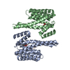
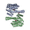
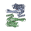
Entry
Database: PDB / ID: 5j31
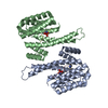
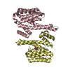
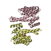
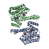
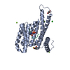
Title
Crystal structure of 14-3-3zeta in complex with an alkyne cross-linked cyclic peptide derived from ExoS
Components
14-3-3 protein zeta/delta
Exoenzyme S
Keywords
SIGNALING PROTEIN / constrained peptides / protein-protein-interaction / alkyne cross-link / 14-3-3 protein zeta
Function / homology
Function and homology information
synaptic target recognition / Golgi reassembly / NOTCH4 Activation and Transmission of Signal to the Nucleus / establishment of Golgi localization / respiratory system process / tube formation / regulation of synapse maturation / Rap1 signalling / negative regulation of protein localization to nucleus / KSRP (KHSRP) binds and destabilizes mRNA ...synaptic target recognition / Golgi reassembly / NOTCH4 Activation and Transmission of Signal to the Nucleus / establishment of Golgi localization / respiratory system process / tube formation / regulation of synapse maturation / Rap1 signalling / negative regulation of protein localization to nucleus / KSRP (KHSRP) binds and destabilizes mRNA / GP1b-IX-V activation signalling / glycosyltransferase activity / Regulation of localization of FOXO transcription factors / Interleukin-3, Interleukin-5 and GM-CSF signaling / phosphoserine residue binding / Activation of BAD and translocation to mitochondria / regulation of ERK1 and ERK2 cascade / SARS-CoV-2 targets host intracellular signalling and regulatory pathways / cellular response to glucose starvation / Chk1/Chk2(Cds1) mediated inactivation of Cyclin B:Cdk1 complex / SARS-CoV-1 targets host intracellular signalling and regulatory pathways / RHO GTPases activate PKNs / negative regulation of TORC1 signaling / nucleotidyltransferase activity / lung development / Transcriptional and post-translational regulation of MITF-M expression and activity / negative regulation of innate immune response / hippocampal mossy fiber to CA3 synapse / GTPase activator activity / TP53 Regulates Metabolic Genes / Translocation of SLC2A4 (GLUT4) to the plasma membrane / protein sequestering activity / Deactivation of the beta-catenin transactivating complex / regulation of protein stability / Negative regulation of NOTCH4 signaling / melanosome / intracellular protein localization / toxin activity / angiogenesis / protein phosphatase binding / blood microparticle / DNA-binding transcription factor binding / vesicle / protein phosphorylation / transmembrane transporter binding / cadherin binding / protein domain specific binding / focal adhesion / ubiquitin protein ligase binding / protein kinase binding / negative regulation of apoptotic process / glutamatergic synapse / negative regulation of transcription by RNA polymerase II / signal transduction / : / RNA binding / extracellular exosome / extracellular region / nucleoplasm / identical protein binding / nucleus / cytosol / cytoplasm Similarity search - Function
Type III secretion system effector protein YopE-like / Virulence factor YopE, GAP domain / Virulence factor YopE, GAP domain superfamily / Yersinia virulence determinant (YopE) / : / ADP ribosyltransferase / ADP-ribosyltransferase exoenzyme / Toxin-related mono-ADP-ribosyltransferase (TR mART) core domain profile. / 14-3-3 domain / Delta-Endotoxin; domain 1 ...Type III secretion system effector protein YopE-like / Virulence factor YopE, GAP domain / Virulence factor YopE, GAP domain superfamily / Yersinia virulence determinant (YopE) / : / ADP ribosyltransferase / ADP-ribosyltransferase exoenzyme / Toxin-related mono-ADP-ribosyltransferase (TR mART) core domain profile. / 14-3-3 domain / Delta-Endotoxin; domain 1 / 14-3-3 proteins signature 2. / 14-3-3 protein, conserved site / 14-3-3 proteins signature 1. / 14-3-3 protein / 14-3-3 homologues / 14-3-3 domain / 14-3-3 domain superfamily / 14-3-3 protein / Up-down Bundle / Mainly Alpha Similarity search - Domain/homology
Resolution: 2.4→47.94 Å / Cor.coef. Fo:Fc: 0.975 / Cor.coef. Fo:Fc free: 0.955 / SU B: 5.801 / SU ML: 0.129 / SU R Cruickshank DPI: 0.175 / Cross valid method: THROUGHOUT / σ(F): 0 / ESU R: 0.175 / ESU R Free: 0.176 Details: HYDROGENS HAVE BEEN ADDED IN THE RIDING POSITIONS U VALUES : REFINED INDIVIDUALLY
Rfactor
Num. reflection
% reflection
Selection details
Rfree
0.2186
2025
5 %
RANDOM
Rwork
0.1631
-
-
-
obs
0.1659
38425
99.96 %
-
Solvent computation
Ion probe radii: 0.8 Å / Shrinkage radii: 0.8 Å / VDW probe radii: 1.2 Å
In the structure databanks used in Yorodumi, some data are registered as the other names, "COVID-19 virus" and "2019-nCoV". Here are the details of the virus and the list of structure data.
Jan 31, 2019. EMDB accession codes are about to change! (news from PDBe EMDB page)
EMDB accession codes are about to change! (news from PDBe EMDB page)
The allocation of 4 digits for EMDB accession codes will soon come to an end. Whilst these codes will remain in use, new EMDB accession codes will include an additional digit and will expand incrementally as the available range of codes is exhausted. The current 4-digit format prefixed with “EMD-” (i.e. EMD-XXXX) will advance to a 5-digit format (i.e. EMD-XXXXX), and so on. It is currently estimated that the 4-digit codes will be depleted around Spring 2019, at which point the 5-digit format will come into force.
The EM Navigator/Yorodumi systems omit the EMD- prefix.
Related info.:Q: What is EMD? / ID/Accession-code notation in Yorodumi/EM Navigator
Yorodumi is a browser for structure data from EMDB, PDB, SASBDB, etc.
This page is also the successor to EM Navigator detail page, and also detail information page/front-end page for Omokage search.
The word "yorodu" (or yorozu) is an old Japanese word meaning "ten thousand". "mi" (miru) is to see.
Related info.:EMDB / PDB / SASBDB / Comparison of 3 databanks / Yorodumi Search / Aug 31, 2016. New EM Navigator & Yorodumi / Yorodumi Papers / Jmol/JSmol / Function and homology information / Changes in new EM Navigator and Yorodumi
 Movie
Movie Controller

 Yorodumi
 Open data
 Basic information
 Components
 Keywords
 Function and homology information
 Homo sapiens (human)
 Pseudomonas aeruginosa (bacteria)
 X-RAY DIFFRACTION /
  Authors
 Citation
 Structure visualization
 Downloads & links
 Other downloads
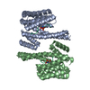


 PDBj
PDBj


 Assembly

 Mass: 122.121 Da / Num. of mol.: 1 / Source method: obtained synthetically / Formula: C7H6O2
 Mass: 18.015 Da / Num. of mol.: 358 / Source method: isolated from a natural source / Formula: H2O
 Sample preparation
 / Beamline: X10SA / Wavelength: 1 Å
 Processing