 Movie
Movie Controller


+ Open data

- Basic information
| Entry | Database: PDB / ID: 2c23 | ||||||
|---|---|---|---|---|---|---|---|
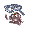
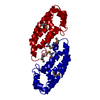
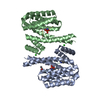
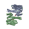
| Title | 14-3-3 Protein Beta (Human) in complex with exoenzyme S peptide | ||||||
 Components Components |
| ||||||
 Keywords Keywords | SIGNALING PROTEIN / 14-3-3 / YWHAB / EXOS / EXOENZYME S / STRUCTURAL GENOMICS / STRUCTURAL GENOMICS CONSORTIUM / ACETYLATION / ALTERNATIVE INITIATION / PHOSPHORYLATION / CELL REGULATOR PROTEIN | ||||||
| Function / homology |  Function and homology information Function and homology informationnegative regulation of cell growth involved in contact inhibition / Tristetraprolin (TTP, ZFP36) binds and destabilizes mRNA / positive regulation of hippo signaling / Butyrate Response Factor 1 (BRF1) binds and destabilizes mRNA / MTOR signalling / ARMS-mediated activation / SHOC2 M1731 mutant abolishes MRAS complex function / Gain-of-function MRAS complexes activate RAF signaling / Rap1 signalling / Signaling by Hippo ...negative regulation of cell growth involved in contact inhibition / Tristetraprolin (TTP, ZFP36) binds and destabilizes mRNA / positive regulation of hippo signaling / Butyrate Response Factor 1 (BRF1) binds and destabilizes mRNA / MTOR signalling / ARMS-mediated activation / SHOC2 M1731 mutant abolishes MRAS complex function / Gain-of-function MRAS complexes activate RAF signaling / Rap1 signalling / Signaling by Hippo / vacuolar membrane / negative regulation of G protein-coupled receptor signaling pathway / negative regulation of protein import into nucleus / Frs2-mediated activation / protein phosphatase inhibitor activity / glycosyltransferase activity / protein kinase inhibitor activity / Regulation of localization of FOXO transcription factors / mTORC1-mediated signalling / phosphoserine residue binding / Activation of BAD and translocation to mitochondria / SARS-CoV-2 targets host intracellular signalling and regulatory pathways / SARS-CoV-1 targets host intracellular signalling and regulatory pathways / RHO GTPases activate PKNs / protein targeting / Chk1/Chk2(Cds1) mediated inactivation of Cyclin B:Cdk1 complex / transcription repressor complex / nucleotidyltransferase activity / Transcriptional and post-translational regulation of MITF-M expression and activity / GTPase activator activity / TP53 Regulates Metabolic Genes / Translocation of SLC2A4 (GLUT4) to the plasma membrane / protein sequestering activity / phosphoprotein binding / RAF activation / Signaling by high-kinase activity BRAF mutants / MAP2K and MAPK activation / histone deacetylase binding / Signaling by RAF1 mutants / Signaling by moderate kinase activity BRAF mutants / Paradoxical activation of RAF signaling by kinase inactive BRAF / Signaling downstream of RAS mutants / Negative regulation of MAPK pathway / Signaling by BRAF and RAF1 fusions / melanosome / intracellular protein localization / toxin activity / cadherin binding / protein domain specific binding / focal adhesion / negative regulation of DNA-templated transcription / protein-containing complex binding / perinuclear region of cytoplasm / enzyme binding / signal transduction / positive regulation of transcription by RNA polymerase II / extracellular exosome / extracellular region / membrane / identical protein binding / nucleus / cytoplasm / cytosol Similarity search - Function | ||||||
| Biological species |  HOMO SAPIENS (human) HOMO SAPIENS (human)  PSEUDOMONAS AERUGINOSA (bacteria) PSEUDOMONAS AERUGINOSA (bacteria) | ||||||
| Method |  X-RAY DIFFRACTION / SYNCHROTRON / MOLECULAR REPLACEMENT / Resolution: 2.65 Å X-RAY DIFFRACTION / SYNCHROTRON / MOLECULAR REPLACEMENT / Resolution: 2.65 Å | ||||||
 Authors Authors | Elkins, J.M. / Schoch, G.A. / Yang, X. / Sundstrom, M. / Arrowsmith, C. / Edwards, A. / Doyle, D.A. | ||||||
 Citation Citation | Journal: Proc.Natl.Acad.Sci.USA / Year: 2006 Title: Structural Basis for Protein-Protein Interactions in the 14-3-3 Protein Family. Authors: Yang, X. / Lee, W.H. / Sobott, F. / Papagrigoriou, E. / Robinson, C.V. / Grossmann, J.G. / Sundstrom, M. / Doyle, D.A. / Elkins, J.M. | ||||||
| History |
|
- Structure visualization
| Structure viewer | Molecule: MolmilJmol/JSmol |
|---|
- Downloads & links
-Download
| PDBx/mmCIF format | 2c23.cif.gz | 58.4 KB | Display | PDBx/mmCIF format |
|---|---|---|---|---|
| PDB format | pdb2c23.ent.gz | 42.7 KB | Display | PDB format |
| PDBx/mmJSON format | 2c23.json.gz | Tree view | PDBx/mmJSON format | |
| Others |  Other downloads Other downloads |
-Validation report
| Arichive directory | https://data.pdbj.org/pub/pdb/validation_reports/c2/2c23ftp://data.pdbj.org/pub/pdb/validation_reports/c2/2c23 | HTTPS FTP |
|---|
-Related structure data
| Related structure data | 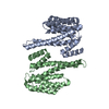 2bq0SC  2br9C  2btpC  2c63C  2c74C S: Starting model for refinement C: citing same article ( |
|---|---|
| Similar structure data |


-Links
 PDBj
PDBj


- Assembly
| Deposited unit | 
| ||||||||
|---|---|---|---|---|---|---|---|---|---|
| 1 | 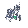
| ||||||||
| Unit cell |
| ||||||||
| Details | THE PROTEIN IS A DIMER IN SOLUTION, BUT SINCE IN THIS ENTRY IT IS IN COMPLEX WITH A PEPTIDE (CHAIN P), THE ENTRY IS MARKED AS TETRAMERIC |
-Components
| #1: Protein | Mass: 28236.627 Da / Num. of mol.: 1 Source method: isolated from a genetically manipulated source Source: (gene. exp.) HOMO SAPIENS (human) / Plasmid: PTVHR21-SGC / Production host: |
|---|---|
| #2: Protein/peptide | Mass: 1116.287 Da / Num. of mol.: 1 / Fragment: 14-3-3 BINDING REGION, RESIDUES 421-431 / Source method: obtained synthetically / Source: (synth.) PSEUDOMONAS AERUGINOSA (bacteria) / References: UniProt: Q51451 |
| Sequence details | RESIDUES 240-245 ARE CLONING ARTEFACT FOR CHAIN A. THE UNIPROT CROSS-REFERENCE GIVEN IN THE DBREF ...RESIDUES 240-245 ARE CLONING ARTEFACT FOR CHAIN A. THE UNIPROT CROSS-REFERENCE GIVEN IN THE DBREF RECORDS BELOW CORRESPOND |
-Experimental details
-Experiment
| Experiment | Method: X-RAY DIFFRACTION / Number of used crystals: 1 |
|---|
- Sample preparation
| Crystal | Density Matthews: 2.81 Å3/Da / Density % sol: 55.9 % |
|---|---|
| Crystal grow | pH: 8 Details: 0.05M MGCL2,0.1M HEPES PH7.5, 30% PEG MME 550, pH 8.00 |
-Data collection
| Diffraction | Mean temperature: 100 K |
|---|---|
| Diffraction source | Source: SYNCHROTRON / Site: SLS  / Beamline: X10SA / Wavelength: 0.99188 / Beamline: X10SA / Wavelength: 0.99188 |
| Detector | Type: ADSC CCD / Detector: CCD / Date: Jul 3, 2005 |
| Radiation | Protocol: SINGLE WAVELENGTH / Monochromatic (M) / Laue (L): M / Scattering type: x-ray |
| Radiation wavelength | Wavelength: 0.99188 Å / Relative weight: 1 |
| Reflection | Resolution: 2.65→49.33 Å / Num. obs: 9558 / % possible obs: 99.9 % / Observed criterion σ(I): 0 / Redundancy: 6.7 % / Rmerge(I) obs: 0.14 / Net I/σ(I): 13.4 |
| Reflection shell | Resolution: 2.65→2.79 Å / Redundancy: 6.8 % / Rmerge(I) obs: 0.43 / Mean I/σ(I) obs: 3 / % possible all: 100 |
- Processing
| Software |
| ||||||||||||||||||||||||||||||||||||||||||||||||||||||||||||||||||||||||||||||||||||||||||||||||||||||||||||||||||||||||||||||||||||||||||||||||||||||||||||||||||||||||||||||||||||||
|---|---|---|---|---|---|---|---|---|---|---|---|---|---|---|---|---|---|---|---|---|---|---|---|---|---|---|---|---|---|---|---|---|---|---|---|---|---|---|---|---|---|---|---|---|---|---|---|---|---|---|---|---|---|---|---|---|---|---|---|---|---|---|---|---|---|---|---|---|---|---|---|---|---|---|---|---|---|---|---|---|---|---|---|---|---|---|---|---|---|---|---|---|---|---|---|---|---|---|---|---|---|---|---|---|---|---|---|---|---|---|---|---|---|---|---|---|---|---|---|---|---|---|---|---|---|---|---|---|---|---|---|---|---|---|---|---|---|---|---|---|---|---|---|---|---|---|---|---|---|---|---|---|---|---|---|---|---|---|---|---|---|---|---|---|---|---|---|---|---|---|---|---|---|---|---|---|---|---|---|---|---|---|---|
| Refinement | Method to determine structure: MOLECULAR REPLACEMENT Starting model: PDB ENTRY 2BQ0 Resolution: 2.65→60.86 Å / Cor.coef. Fo:Fc: 0.944 / Cor.coef. Fo:Fc free: 0.898 / SU B: 13.677 / SU ML: 0.28 / Cross valid method: THROUGHOUT / ESU R: 0.631 / ESU R Free: 0.35 / Stereochemistry target values: MAXIMUM LIKELIHOOD / Details: HYDROGENS HAVE BEEN ADDED IN THE RIDING POSITIONS.
| ||||||||||||||||||||||||||||||||||||||||||||||||||||||||||||||||||||||||||||||||||||||||||||||||||||||||||||||||||||||||||||||||||||||||||||||||||||||||||||||||||||||||||||||||||||||
| Solvent computation | Ion probe radii: 0.8 Å / Shrinkage radii: 0.8 Å / VDW probe radii: 1.2 Å / Solvent model: MASK | ||||||||||||||||||||||||||||||||||||||||||||||||||||||||||||||||||||||||||||||||||||||||||||||||||||||||||||||||||||||||||||||||||||||||||||||||||||||||||||||||||||||||||||||||||||||
| Displacement parameters | Biso mean: 61.64 Å2
| ||||||||||||||||||||||||||||||||||||||||||||||||||||||||||||||||||||||||||||||||||||||||||||||||||||||||||||||||||||||||||||||||||||||||||||||||||||||||||||||||||||||||||||||||||||||
| Refinement step | Cycle: LAST / Resolution: 2.65→60.86 Å
| ||||||||||||||||||||||||||||||||||||||||||||||||||||||||||||||||||||||||||||||||||||||||||||||||||||||||||||||||||||||||||||||||||||||||||||||||||||||||||||||||||||||||||||||||||||||
| Refine LS restraints |
|