 Movie
Movie Controller

[English] 日本語
 Yorodumi
- PDB-3o8i: Structure of 14-3-3 isoform sigma in complex with a C-Raf1 peptid... -
+ Open data

- Basic information
| Entry | Database: PDB / ID: 3o8i | ||||||
|---|---|---|---|---|---|---|---|
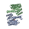
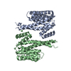
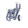
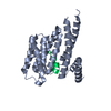
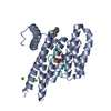
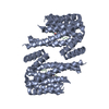
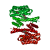
| Title | Structure of 14-3-3 isoform sigma in complex with a C-Raf1 peptide and a stabilizing small molecule fragment | ||||||
 Components Components |
| ||||||
 Keywords Keywords | PEPTIDE BINDING PROTEIN / Protein-Protein Complex / 14-3-3 / Protein-protein interaction / Phosphorylation | ||||||
| Function / homology |  Function and homology information Function and homology informationdeath-inducing signaling complex assembly / intermediate filament cytoskeleton organization / regulation of Rho protein signal transduction / SHOC2 M1731 mutant abolishes MRAS complex function / Gain-of-function MRAS complexes activate RAF signaling / Rap1 signalling / type B pancreatic cell proliferation / insulin secretion involved in cellular response to glucose stimulus / Negative feedback regulation of MAPK pathway / IFNG signaling activates MAPKs ...death-inducing signaling complex assembly / intermediate filament cytoskeleton organization / regulation of Rho protein signal transduction / SHOC2 M1731 mutant abolishes MRAS complex function / Gain-of-function MRAS complexes activate RAF signaling / Rap1 signalling / type B pancreatic cell proliferation / insulin secretion involved in cellular response to glucose stimulus / Negative feedback regulation of MAPK pathway / IFNG signaling activates MAPKs / GP1b-IX-V activation signalling / regulation of epidermal cell division / protein kinase C inhibitor activity / positive regulation of epidermal cell differentiation / keratinocyte development / keratinization / ERBB2-ERBB3 signaling pathway / neurotrophin TRK receptor signaling pathway / face development / regulation of cell-cell adhesion / thyroid gland development / pseudopodium / establishment of skin barrier / regulation of cell differentiation / Regulation of localization of FOXO transcription factors / keratinocyte proliferation / somatic stem cell population maintenance / extrinsic apoptotic signaling pathway via death domain receptors / positive regulation of peptidyl-serine phosphorylation / negative regulation of keratinocyte proliferation / Activation of BAD and translocation to mitochondria / phosphoserine residue binding / MAP kinase kinase kinase activity / type II interferon-mediated signaling pathway / cAMP/PKA signal transduction / negative regulation of protein localization to plasma membrane / SARS-CoV-2 targets host intracellular signalling and regulatory pathways / Schwann cell development / negative regulation of protein kinase activity / negative regulation of stem cell proliferation / negative regulation of extrinsic apoptotic signaling pathway via death domain receptors / negative regulation of protein-containing complex assembly / SARS-CoV-1 targets host intracellular signalling and regulatory pathways / RHO GTPases activate PKNs / Chk1/Chk2(Cds1) mediated inactivation of Cyclin B:Cdk1 complex / positive regulation of protein localization / response to muscle stretch / myelination / protein export from nucleus / insulin-like growth factor receptor signaling pathway / CD209 (DC-SIGN) signaling / TP53 Regulates Transcription of Genes Involved in G2 Cell Cycle Arrest / negative regulation of innate immune response / positive regulation of cell adhesion / release of cytochrome c from mitochondria / positive regulation of protein export from nucleus / thymus development / stem cell proliferation / adenylate cyclase activator activity / TP53 Regulates Metabolic Genes / Translocation of SLC2A4 (GLUT4) to the plasma membrane / wound healing / protein sequestering activity / RAF activation / Signaling by high-kinase activity BRAF mutants / MAP2K and MAPK activation / intrinsic apoptotic signaling pathway in response to DNA damage / Stimuli-sensing channels / Signaling by RAF1 mutants / Signaling by moderate kinase activity BRAF mutants / Paradoxical activation of RAF signaling by kinase inactive BRAF / Signaling downstream of RAS mutants / Negative regulation of MAPK pathway / insulin receptor signaling pathway / Signaling by BRAF and RAF1 fusions / intracellular protein localization / MAPK cascade / regulation of protein localization / positive regulation of cell growth / sperm midpiece / regulation of apoptotic process / protein phosphorylation / protein kinase activity / positive regulation of MAPK cascade / mitochondrial outer membrane / non-specific serine/threonine protein kinase / regulation of cell cycle / cadherin binding / negative regulation of cell population proliferation / protein serine kinase activity / protein serine/threonine kinase activity / apoptotic process / protein kinase binding / negative regulation of apoptotic process / enzyme binding / negative regulation of transcription by RNA polymerase II / Golgi apparatus / signal transduction / positive regulation of transcription by RNA polymerase II / mitochondrion Similarity search - Function | ||||||
| Biological species |  Homo sapiens (human) Homo sapiens (human) | ||||||
| Method |  X-RAY DIFFRACTION / SYNCHROTRON / MOLECULAR REPLACEMENT / molecular replacement / Resolution: 2 Å X-RAY DIFFRACTION / SYNCHROTRON / MOLECULAR REPLACEMENT / molecular replacement / Resolution: 2 Å | ||||||
 Authors Authors | Ottmann, C. / Rose, R. / Kaiser, M. / Kuhenne, P. | ||||||
 Citation Citation | Journal: Mol.Cell.Biol. / Year: 2010 Title: Impaired Binding of 14-3-3 to C-RAF in Noonan Syndrome Suggests New Approaches in Diseases with Increased Ras Signaling Authors: Molzan, M. / Schumacher, B. / Ottmann, C. / Baljuls, A. / Polzien, L. / Weyand, M. / Thiel, P. / Rose, R. / Rose, M. / Kuhenne, P. / Kaiser, M. / Rapp, U.R. / Kuhlmann, J. / Ottmann, C. | ||||||
| History |
|
- Structure visualization
| Structure viewer | Molecule: MolmilJmol/JSmol |
|---|
- Downloads & links
-Download
| PDBx/mmCIF format | 3o8i.cif.gz | 112.8 KB | Display | PDBx/mmCIF format |
|---|---|---|---|---|
| PDB format | pdb3o8i.ent.gz | 86.7 KB | Display | PDB format |
| PDBx/mmJSON format | 3o8i.json.gz | Tree view | PDBx/mmJSON format | |
| Others |  Other downloads Other downloads |
-Validation report
| Arichive directory | https://data.pdbj.org/pub/pdb/validation_reports/o8/3o8iftp://data.pdbj.org/pub/pdb/validation_reports/o8/3o8i | HTTPS FTP |
|---|
-Related structure data
| Related structure data | 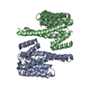 3cu8C 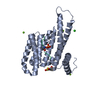 3iqjC 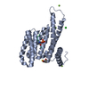 3iquSC 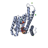 3iqvC 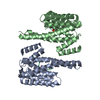 3nkxC C: citing same article ( S: Starting model for refinement |
|---|---|
| Similar structure data |


-Links
 PDBj
PDBj


- Assembly
| Deposited unit | 
| ||||||||
|---|---|---|---|---|---|---|---|---|---|
| 1 | 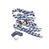
| ||||||||
| Unit cell |
|
-Components
| #1: Protein | Mass: 26818.242 Da / Num. of mol.: 1 / Fragment: C-terminal deletion Source method: isolated from a genetically manipulated source Source: (gene. exp.) Homo sapiens (human) / Gene: SFN, HME1 / Plasmid: pProEX-Htb / Production host:  |
|---|---|
| #2: Protein/peptide | Mass: 1208.175 Da / Num. of mol.: 1 / Source method: obtained synthetically / Details: Synthesized Phospho-Peptide / References: UniProt: P04049 |
| #3: Chemical | ChemComp-M1T /   Mass: 162.184 Da / Num. of mol.: 1 / Source method: obtained synthetically / Formula: C7H14O4 Mass: 162.184 Da / Num. of mol.: 1 / Source method: obtained synthetically / Formula: C7H14O4 |
| #4: Water | ChemComp-HOH /  Mass: 18.015 Da / Num. of mol.: 163 / Source method: isolated from a natural source / Formula: H2O Mass: 18.015 Da / Num. of mol.: 163 / Source method: isolated from a natural source / Formula: H2O |
| Has protein modification | Y |
-Experimental details
-Experiment
| Experiment | Method: X-RAY DIFFRACTION / Number of used crystals: 1 |
|---|
- Sample preparation
| Crystal | Density Matthews: 2.77 Å3/Da / Density % sol: 55.61 % |
|---|---|
| Crystal grow | Temperature: 277.15 K / Method: vapor diffusion, hanging drop / pH: 7.5 Details: 0.095M HEPES Na, 26.6%(v/v) PEG400, 0.19M CaCl2, 5%(v/v) Glycerol, pH 7.5, VAPOR DIFFUSION, HANGING DROP, temperature 277.15K |
-Data collection
| Diffraction | Mean temperature: 100 K | ||||||||||||||||||||||||||||||||||||||||||||||||||||||||||||||||||||||||||||||||||||||||||||||||||||
|---|---|---|---|---|---|---|---|---|---|---|---|---|---|---|---|---|---|---|---|---|---|---|---|---|---|---|---|---|---|---|---|---|---|---|---|---|---|---|---|---|---|---|---|---|---|---|---|---|---|---|---|---|---|---|---|---|---|---|---|---|---|---|---|---|---|---|---|---|---|---|---|---|---|---|---|---|---|---|---|---|---|---|---|---|---|---|---|---|---|---|---|---|---|---|---|---|---|---|---|---|---|
| Diffraction source | Source: SYNCHROTRON / Site: SLS  / Beamline: X10SA / Wavelength: 0.99986 Å / Beamline: X10SA / Wavelength: 0.99986 Å | ||||||||||||||||||||||||||||||||||||||||||||||||||||||||||||||||||||||||||||||||||||||||||||||||||||
| Detector | Type: MARMOSAIC 225 mm CCD / Detector: CCD / Date: Aug 11, 2009 | ||||||||||||||||||||||||||||||||||||||||||||||||||||||||||||||||||||||||||||||||||||||||||||||||||||
| Radiation | Monochromator: Si(111) monochromator (horizontal) and Dynamically bendable mirror (vertical) Protocol: SINGLE WAVELENGTH / Monochromatic (M) / Laue (L): M / Scattering type: x-ray | ||||||||||||||||||||||||||||||||||||||||||||||||||||||||||||||||||||||||||||||||||||||||||||||||||||
| Radiation wavelength | Wavelength: 0.99986 Å / Relative weight: 1 | ||||||||||||||||||||||||||||||||||||||||||||||||||||||||||||||||||||||||||||||||||||||||||||||||||||
| Reflection | Resolution: 2→20 Å / Num. obs: 21324 / % possible obs: 99.5 % / Observed criterion σ(I): -3 / Biso Wilson estimate: 33.855 Å2 / Rmerge(I) obs: 0.072 / Net I/σ(I): 14.37 | ||||||||||||||||||||||||||||||||||||||||||||||||||||||||||||||||||||||||||||||||||||||||||||||||||||
| Reflection shell |
|
-Phasing
| Phasing | Method: molecular replacement |
|---|
- Processing
| Software |
| ||||||||||||||||||||||||||||||||||||||||||||||||||||||||||||||||||||||
|---|---|---|---|---|---|---|---|---|---|---|---|---|---|---|---|---|---|---|---|---|---|---|---|---|---|---|---|---|---|---|---|---|---|---|---|---|---|---|---|---|---|---|---|---|---|---|---|---|---|---|---|---|---|---|---|---|---|---|---|---|---|---|---|---|---|---|---|---|---|---|---|
| Refinement | Method to determine structure: MOLECULAR REPLACEMENT Starting model: 3IQU Resolution: 2→19.58 Å / Cor.coef. Fo:Fc: 0.95 / Cor.coef. Fo:Fc free: 0.925 / Occupancy max: 1 / Occupancy min: 0.3 / Cross valid method: THROUGHOUT / σ(F): 0 / ESU R Free: 0.154 / Stereochemistry target values: MAXIMUM LIKELIHOOD Details: HYDROGENS HAVE BEEN ADDED IN THE RIDING POSITIONS U VALUES REFINED INDIVIDUALLY
| ||||||||||||||||||||||||||||||||||||||||||||||||||||||||||||||||||||||
| Solvent computation | Ion probe radii: 0.8 Å / Shrinkage radii: 0.8 Å / VDW probe radii: 1.4 Å / Solvent model: MASK | ||||||||||||||||||||||||||||||||||||||||||||||||||||||||||||||||||||||
| Displacement parameters | Biso max: 73.42 Å2 / Biso mean: 29.5154 Å2 / Biso min: 8.35 Å2
| ||||||||||||||||||||||||||||||||||||||||||||||||||||||||||||||||||||||
| Refinement step | Cycle: LAST / Resolution: 2→19.58 Å
| ||||||||||||||||||||||||||||||||||||||||||||||||||||||||||||||||||||||
| Refine LS restraints |
| ||||||||||||||||||||||||||||||||||||||||||||||||||||||||||||||||||||||
| LS refinement shell | Resolution: 2→2.051 Å / Total num. of bins used: 20
|
