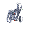
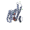
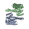
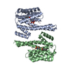
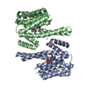
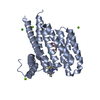
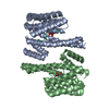
Entry Database : PDB / ID : 3cu8Title Impaired binding of 14-3-3 to Raf1 is linked to Noonan and LEOPARD syndrome 14-3-3 protein zeta/delta RAF proto-oncogene serine/threonine-protein kinase Keywords / / / / / / / / Function / homology Function Domain/homology Component
/ / / / / / / / / / / / / / / / / / / / / / / / / / / / / / / / / / / / / / / / / / / / / / / / / / / / / / / / / / / / / / / / / / / / / / / / / / / / / / / / / / / / / / / / / / / / / / / / / / / / / / / / / / / / / / / / / / / / / / / / / / / / / / / / / / / / / / / / / / / / / / / / / / / / / / / Biological species Homo sapiens (human)Method / / / Resolution : 2.4 Å Authors Schumacher, B. / Weyand, M. / Kuhlmann, J. / Ottmann, C. Journal : Mol. Cell. Biol. / Year : 2010Title : Impaired binding of 14-3-3 to C-RAF in Noonan syndrome suggests new approaches in diseases with increased Ras signaling.Authors : Molzan, M. / Schumacher, B. / Ottmann, C. / Baljuls, A. / Polzien, L. / Weyand, M. / Thiel, P. / Rose, R. / Rose, M. / Kuhenne, P. / Kaiser, M. / Rapp, U.R. / Kuhlmann, J. / Ottmann, C. History Deposition Apr 16, 2008 Deposition site / Processing site Revision 1.0 May 5, 2009 Provider / Type Revision 1.1 Jul 13, 2011 Group / Version format complianceRevision 1.2 Jun 7, 2017 Group Revision 1.3 Oct 25, 2017 Group / Category Revision 1.4 Nov 13, 2024 Group Data collection / Database references ... Data collection / Database references / Derived calculations / Structure summary Category chem_comp_atom / chem_comp_bond ... chem_comp_atom / chem_comp_bond / database_2 / pdbx_entry_details / pdbx_modification_feature / struct_conn / struct_site Item _database_2.pdbx_DOI / _database_2.pdbx_database_accession ... _database_2.pdbx_DOI / _database_2.pdbx_database_accession / _struct_conn.pdbx_leaving_atom_flag / _struct_site.pdbx_auth_asym_id / _struct_site.pdbx_auth_comp_id / _struct_site.pdbx_auth_seq_id
Show all Show less
 Movie
Movie Controller

 Yorodumi
 Open data
 Basic information
 Components
 Keywords
 Function and homology information
 Homo sapiens (human)
 X-RAY DIFFRACTION /
  Authors
 Citation
 Structure visualization
 Downloads & links
 Other downloads
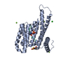


 PDBj
PDBj


 Assembly


 Mass: 24.305 Da / Num. of mol.: 2 / Source method: obtained synthetically / Formula: Mg
 Mass: 74.079 Da / Num. of mol.: 1 / Source method: obtained synthetically / Formula: C3H6O2
 Mass: 18.015 Da / Num. of mol.: 196 / Source method: isolated from a natural source / Formula: H2O
 Sample preparation
 / Beamline: X10SA / Wavelength: 0.97809 Å
 Processing