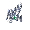
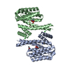
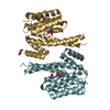
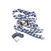
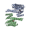
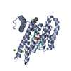
[English] 日本語
Yorodumi
- PDB-6ejl: Structure of 14-3-3 zeta in complex with ASK1 14-3-3 binding motif -

+
Open data


ID or keywords:

Loading...

-
Basic information

Entry
Database: PDB / ID: 6ejl
TitleStructure of 14-3-3 zeta in complex with ASK1 14-3-3 binding motif
Components
  • 14-3-3 protein zeta/delta
  • Mitogen-activated protein kinase kinase kinase 5
KeywordsSIGNALING PROTEIN / 14-3-3 protein / ASK1 / Apoptosis signal-regulating kinase 1
Function / homology
Function and homology information


cellular response to reactive nitrogen species / neuron intrinsic apoptotic signaling pathway in response to oxidative stress / IRE1-TRAF2-ASK1 complex / Golgi reassembly / synaptic target recognition / protein kinase complex / mitogen-activated protein kinase kinase kinase / programmed necrotic cell death / respiratory system process / NOTCH4 Activation and Transmission of Signal to the Nucleus ...cellular response to reactive nitrogen species / neuron intrinsic apoptotic signaling pathway in response to oxidative stress / IRE1-TRAF2-ASK1 complex / Golgi reassembly / synaptic target recognition / protein kinase complex / mitogen-activated protein kinase kinase kinase / programmed necrotic cell death / respiratory system process / NOTCH4 Activation and Transmission of Signal to the Nucleus / regulation of synapse maturation / JUN kinase kinase kinase activity / establishment of Golgi localization / tube formation / endothelial cell apoptotic process / Rap1 signalling / negative regulation of protein localization to nucleus / positive regulation of p38MAPK cascade / KSRP (KHSRP) binds and destabilizes mRNA / intrinsic apoptotic signaling pathway in response to oxidative stress / GP1b-IX-V activation signalling / positive regulation of cardiac muscle cell apoptotic process / p38MAPK cascade / positive regulation of cysteine-type endopeptidase activity involved in apoptotic process / Regulation of localization of FOXO transcription factors / Interleukin-3, Interleukin-5 and GM-CSF signaling / phosphoserine residue binding / MAP kinase kinase kinase activity / intrinsic apoptotic signaling pathway in response to endoplasmic reticulum stress / Activation of BAD and translocation to mitochondria / positive regulation of protein kinase activity / positive regulation of myoblast differentiation / protein targeting / SARS-CoV-2 targets host intracellular signalling and regulatory pathways / cellular response to glucose starvation / Chk1/Chk2(Cds1) mediated inactivation of Cyclin B:Cdk1 complex / SARS-CoV-1 targets host intracellular signalling and regulatory pathways / stress-activated MAPK cascade / positive regulation of JUN kinase activity / RHO GTPases activate PKNs / JNK cascade / negative regulation of TORC1 signaling / positive regulation of vascular associated smooth muscle cell proliferation / protein sequestering activity / ERK1 and ERK2 cascade / cellular response to amino acid starvation / negative regulation of innate immune response / hippocampal mossy fiber to CA3 synapse / response to endoplasmic reticulum stress / regulation of ERK1 and ERK2 cascade / response to ischemia / Translocation of SLC2A4 (GLUT4) to the plasma membrane / Deactivation of the beta-catenin transactivating complex / TP53 Regulates Metabolic Genes / apoptotic signaling pathway / positive regulation of JNK cascade / Negative regulation of NOTCH4 signaling / lung development / regulation of protein stability / cellular response to hydrogen peroxide / MAPK cascade / cellular senescence / melanosome / cellular response to tumor necrosis factor / protein phosphatase binding / angiogenesis / Oxidative Stress Induced Senescence / DNA-binding transcription factor binding / neuron apoptotic process / vesicle / transmembrane transporter binding / blood microparticle / protein kinase activity / cadherin binding / positive regulation of apoptotic process / protein domain specific binding / external side of plasma membrane / protein phosphorylation / innate immune response / protein serine kinase activity / focal adhesion / protein serine/threonine kinase activity / glutamatergic synapse / ubiquitin protein ligase binding / negative regulation of apoptotic process / protein kinase binding / positive regulation of DNA-templated transcription / negative regulation of transcription by RNA polymerase II / magnesium ion binding / signal transduction / protein homodimerization activity / protein-containing complex / RNA binding / extracellular space / extracellular exosome / nucleoplasm / ATP binding / identical protein binding / nucleus / cytoplasm
Similarity search - Function
MAP3K, deoxyribohydrolase domain / MAP3K, HisK-N-like globin domain / HisK-N-like globin domain of the ASK signalosome / Deoxyribohydrolase (DRHyd) domain of the ASK signalosome / MAP3K, TRAFs-binding domain / MAP3K, PH domain / MAP3K TRAFs-binding domain / ASK kinase PH domain / 14-3-3 domain / Delta-Endotoxin; domain 1 ...MAP3K, deoxyribohydrolase domain / MAP3K, HisK-N-like globin domain / HisK-N-like globin domain of the ASK signalosome / Deoxyribohydrolase (DRHyd) domain of the ASK signalosome / MAP3K, TRAFs-binding domain / MAP3K, PH domain / MAP3K TRAFs-binding domain / ASK kinase PH domain / 14-3-3 domain / Delta-Endotoxin; domain 1 / 14-3-3 proteins signature 2. / 14-3-3 protein, conserved site / 14-3-3 proteins signature 1. / 14-3-3 protein / 14-3-3 homologues / 14-3-3 domain / 14-3-3 domain superfamily / 14-3-3 protein / Sterile alpha motif/pointed domain superfamily / Serine/threonine-protein kinase, active site / Serine/Threonine protein kinases active-site signature. / Protein kinase domain / Serine/Threonine protein kinases, catalytic domain / Protein kinase, ATP binding site / Protein kinases ATP-binding region signature. / Protein kinase domain profile. / Protein kinase domain / Protein kinase-like domain superfamily / Up-down Bundle / Mainly Alpha
Similarity search - Domain/homology
14-3-3 protein zeta/delta / Mitogen-activated protein kinase kinase kinase 5
Similarity search - Component
Biological speciesHomo sapiens (human)
MethodX-RAY DIFFRACTION / MOLECULAR REPLACEMENT / Resolution: 2.382 Å
AuthorsPetrvalska, O. / Lentini Santo, D. / Obsilova, V. / Obsil, T.
Funding support Czech Republic, 1items
OrganizationGrant numberCountry
European Commission675179 - TASPPI - H2020-MSCA-ITN-2015 Czech Republic
CitationJournal: To Be Published
Title: Crystal structure of 14-3-3 zeta in complex with ASK1 14-3-3 binding motif
Authors: Petrvalska, O. / Lentini Santo, D. / Obsilova, V. / Obsil, T.
History
DepositionSep 21, 2017Deposition site: PDBE / Processing site: PDBE
Revision 1.0Nov 8, 2017Provider: repository / Type: Initial release
Revision 1.1Mar 7, 2018Group: Data collection / Category: diffrn_source / Item: _diffrn_source.source
Revision 1.2Nov 6, 2019Group: Data collection / Category: diffrn_detector / Item: _diffrn_detector.detector
Revision 1.3Jan 17, 2024Group: Data collection / Database references / Refinement description
Category: chem_comp_atom / chem_comp_bond ...chem_comp_atom / chem_comp_bond / database_2 / pdbx_initial_refinement_model
Item: _database_2.pdbx_DOI / _database_2.pdbx_database_accession

-
Structure visualization

Structure viewerMolecule:
MolmilJmol/JSmol

Downloads & links

-
Assembly

Deposited unit
A: 14-3-3 protein zeta/delta
B: 14-3-3 protein zeta/delta
C: Mitogen-activated protein kinase kinase kinase 5
D: Mitogen-activated protein kinase kinase kinase 5


Theoretical massNumber of molelcules
Total (without water)55,0964
Polymers55,0964
Non-polymers00
Water2,648147
1


  • Idetical with deposited unit
  • defined by author&software
  • Evidence: gel filtration
TypeNameSymmetry operationNumber
identity operation1_555x,y,z1
Buried area4080 Å2
ΔGint-34 kcal/mol
Surface area21510 Å2
MethodPISA
Unit cell
Length a, b, c (Å)74.980, 93.427, 68.491
Angle α, β, γ (deg.)90.00, 99.74, 90.00
Int Tables number5
Space group name H-MC121
Components on special symmetry positions
IDModelComponents
11A-370-

HOH

-
Components

#1: Protein 14-3-3 protein zeta/delta / Protein kinase C inhibitor protein 1 / KCIP-1


Mass: 26599.041 Da / Num. of mol.: 2
Source method: isolated from a genetically manipulated source
Source: (gene. exp.) Homo sapiens (human) / Gene: YWHAZ / Production host: Escherichia coli BL21(DE3) (bacteria) / References: UniProt: P63104
#2: Protein/peptide Mitogen-activated protein kinase kinase kinase 5 / Apoptosis signal-regulating kinase 1 / ASK-1 / MAPK/ERK kinase kinase 5 / MEKK 5


Mass: 949.020 Da / Num. of mol.: 2 / Fragment: UNP residues 963-970 / Source method: obtained synthetically / Source: (synth.) Homo sapiens (human)
References: UniProt: Q99683, mitogen-activated protein kinase kinase kinase
#3: Water ChemComp-HOH / water


Mass: 18.015 Da / Num. of mol.: 147 / Source method: isolated from a natural source / Formula: H2O

-
Experimental details

-
Experiment

ExperimentMethod: X-RAY DIFFRACTION / Number of used crystals: 1

-
Sample preparation

CrystalDensity Matthews: 2.15 Å3/Da / Density % sol: 42.68 %
Crystal growTemperature: 291 K / Method: vapor diffusion, hanging drop / pH: 8.5 / Details: Morpheus HT-96, condition H12

-
Data collection

DiffractionMean temperature: 100 K
Diffraction sourceSource: LIQUID ANODE / Type: Excillum MetalJet D2+ 70 kV / Wavelength: 1.3418 Å
DetectorType: Bruker PHOTON II / Detector: PIXEL / Date: Jun 10, 2016
RadiationProtocol: SINGLE WAVELENGTH / Monochromatic (M) / Laue (L): M / Scattering type: x-ray
Radiation wavelengthWavelength: 1.3418 Å / Relative weight: 1
ReflectionResolution: 2.38→31.01 Å / Num. obs: 18601 / % possible obs: 99.4 % / Redundancy: 6.96 % / CC1/2: 0.999 / Rmerge(I) obs: 0.077 / Net I/σ(I): 20.57
Reflection shellResolution: 2.38→2.53 Å / Redundancy: 6.89 % / Rmerge(I) obs: 0.277 / Mean I/σ(I) obs: 7.15 / Num. unique obs: 2937 / CC1/2: 0.976 / % possible all: 97.8

-
Processing

Software
NameVersionClassification
PHENIX(1.11.1_2575: ???)refinement
XDSdata reduction
XDSdata scaling
MOLREPphasing
RefinementMethod to determine structure: MOLECULAR REPLACEMENT
Starting model: 1QJB
Resolution: 2.382→30.322 Å / SU ML: 0.23 / Cross valid method: FREE R-VALUE / σ(F): 1.36 / Phase error: 23.16
RfactorNum. reflection% reflection
Rfree0.2154 928 5 %
Rwork0.1956 --
obs0.1966 18553 99.4 %
Solvent computationShrinkage radii: 0.9 Å / VDW probe radii: 1.11 Å
Refinement stepCycle: LAST / Resolution: 2.382→30.322 Å
ProteinNucleic acidLigandSolventTotal
Num. atoms3662 0 0 147 3809
Refine LS restraints
Refine-IDTypeDev idealNumber
X-RAY DIFFRACTIONf_bond_d0.0083711
X-RAY DIFFRACTIONf_angle_d0.8945000
X-RAY DIFFRACTIONf_dihedral_angle_d25.0611404
X-RAY DIFFRACTIONf_chiral_restr0.042565
X-RAY DIFFRACTIONf_plane_restr0.005639
LS refinement shell
Resolution (Å)Rfactor RfreeNum. reflection RfreeRfactor RworkNum. reflection RworkRefine-ID% reflection obs (%)
2.3822-2.50770.27321300.22472463X-RAY DIFFRACTION98
2.5077-2.66470.25331310.2342490X-RAY DIFFRACTION100
2.6647-2.87030.26771330.23412539X-RAY DIFFRACTION100
2.8703-3.15890.27221330.22582519X-RAY DIFFRACTION100
3.1589-3.61540.21071330.19362517X-RAY DIFFRACTION100
3.6154-4.55250.16551330.15982535X-RAY DIFFRACTION100
4.5525-30.32460.19191350.18022562X-RAY DIFFRACTION99

+
About Yorodumi

-
News

-
Feb 9, 2022. New format data for meta-information of EMDB entries

New format data for meta-information of EMDB entries

  • Version 3 of the EMDB header file is now the official format.
  • The previous official version 1.9 will be removed from the archive.

Related info.:EMDB header

External links:wwPDB to switch to version 3 of the EMDB data model

-
Aug 12, 2020. Covid-19 info

Covid-19 info

URL: https://pdbjlvh1.pdbj.org/emnavi/covid19.php

New page: Covid-19 featured information page in EM Navigator.

Related info.:Covid-19 info / Mar 5, 2020. Novel coronavirus structure data

+
Mar 5, 2020. Novel coronavirus structure data

Novel coronavirus structure data

Related info.:Yorodumi Speices / Aug 12, 2020. Covid-19 info

External links:COVID-19 featured content - PDBj / Molecule of the Month (242):Coronavirus Proteases

+
Jan 31, 2019. EMDB accession codes are about to change! (news from PDBe EMDB page)

EMDB accession codes are about to change! (news from PDBe EMDB page)

  • The allocation of 4 digits for EMDB accession codes will soon come to an end. Whilst these codes will remain in use, new EMDB accession codes will include an additional digit and will expand incrementally as the available range of codes is exhausted. The current 4-digit format prefixed with “EMD-” (i.e. EMD-XXXX) will advance to a 5-digit format (i.e. EMD-XXXXX), and so on. It is currently estimated that the 4-digit codes will be depleted around Spring 2019, at which point the 5-digit format will come into force.
  • The EM Navigator/Yorodumi systems omit the EMD- prefix.

Related info.:Q: What is EMD? / ID/Accession-code notation in Yorodumi/EM Navigator

External links:EMDB Accession Codes are Changing Soon! / Contact to PDBj

+
Jul 12, 2017. Major update of PDB

Major update of PDB

  • wwPDB released updated PDB data conforming to the new PDBx/mmCIF dictionary.
  • This is a major update changing the version number from 4 to 5, and with Remediation, in which all the entries are updated.
  • In this update, many items about electron microscopy experimental information are reorganized (e.g. em_software).
  • Now, EM Navigator and Yorodumi are based on the updated data.

External links:wwPDB Remediation / Enriched Model Files Conforming to OneDep Data Standards Now Available in the PDB FTP Archive

-
Yorodumi

Thousand views of thousand structures

  • Yorodumi is a browser for structure data from EMDB, PDB, SASBDB, etc.
  • This page is also the successor to EM Navigator detail page, and also detail information page/front-end page for Omokage search.
  • The word "yorodu" (or yorozu) is an old Japanese word meaning "ten thousand". "mi" (miru) is to see.

Related info.:EMDB / PDB / SASBDB / Comparison of 3 databanks / Yorodumi Search / Aug 31, 2016. New EM Navigator & Yorodumi / Yorodumi Papers / Jmol/JSmol / Function and homology information / Changes in new EM Navigator and Yorodumi

Read more