 Movie
Movie Controller


+ Open data

- Basic information
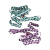
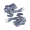
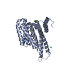
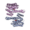
| Entry | Database: PDB / ID: 6byl | ||||||
|---|---|---|---|---|---|---|---|
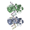
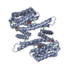
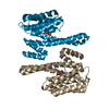
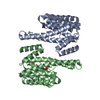
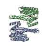
| Title | Structure of 14-3-3 gamma bound to O-GlcNAcylated thr peptide | ||||||
 Components Components |
| ||||||
 Keywords Keywords | PEPTIDE BINDING PROTEIN / 14-3-3 / O-glcNAc | ||||||
| Function / homology |  Function and homology information Function and homology informationpositive regulation of cell-cell adhesion / phosphorylation-dependent protein binding / positive regulation of T cell mediated immune response to tumor cell / regulation of neuron differentiation / protein kinase C inhibitor activity / Regulation of localization of FOXO transcription factors / Activation of BAD and translocation to mitochondria / regulation of signal transduction / SARS-CoV-2 targets host intracellular signalling and regulatory pathways / negative regulation of protein kinase activity ...positive regulation of cell-cell adhesion / phosphorylation-dependent protein binding / positive regulation of T cell mediated immune response to tumor cell / regulation of neuron differentiation / protein kinase C inhibitor activity / Regulation of localization of FOXO transcription factors / Activation of BAD and translocation to mitochondria / regulation of signal transduction / SARS-CoV-2 targets host intracellular signalling and regulatory pathways / negative regulation of protein kinase activity / cellular response to glucose starvation / SARS-CoV-1 targets host intracellular signalling and regulatory pathways / protein targeting / RHO GTPases activate PKNs / Chk1/Chk2(Cds1) mediated inactivation of Cyclin B:Cdk1 complex / insulin-like growth factor receptor binding / negative regulation of TORC1 signaling / Loss of Nlp from mitotic centrosomes / Loss of proteins required for interphase microtubule organization from the centrosome / Transcriptional and post-translational regulation of MITF-M expression and activity / Recruitment of mitotic centrosome proteins and complexes / Recruitment of NuMA to mitotic centrosomes / Anchoring of the basal body to the plasma membrane / AURKA Activation by TPX2 / protein kinase C binding / TP53 Regulates Metabolic Genes / Translocation of SLC2A4 (GLUT4) to the plasma membrane / protein sequestering activity / receptor tyrosine kinase binding / regulation of synaptic plasticity / positive regulation of T cell activation / cellular response to insulin stimulus / intracellular protein localization / Regulation of PLK1 Activity at G2/M Transition / regulation of protein localization / presynapse / mitochondrial matrix / protein domain specific binding / focal adhesion / signal transduction / RNA binding / extracellular exosome / membrane / identical protein binding / nucleus / cytoplasm / cytosol Similarity search - Function | ||||||
| Biological species |  Homo sapiens (human) Homo sapiens (human)synthetic construct (others) | ||||||
| Method |  X-RAY DIFFRACTION / SYNCHROTRON / MOLECULAR REPLACEMENT / molecular replacement / Resolution: 3.35 Å X-RAY DIFFRACTION / SYNCHROTRON / MOLECULAR REPLACEMENT / molecular replacement / Resolution: 3.35 Å | ||||||
 Authors Authors | Schumacher, M.A. | ||||||
 Citation Citation | Journal: Proc.Natl.Acad.Sci.USA / Year: 2018 Title: Structural basis of O-GlcNAc recognition by mammalian 14-3-3 proteins. Authors: Toleman, C.A. / Schumacher, M.A. / Yu, S.H. / Zeng, W. / Cox, N.J. / Smith, T.J. / Soderblom, E.J. / Wands, A.M. / Kohler, J.J. / Boyce, M. | ||||||
| History |
|
- Structure visualization
| Structure viewer | Molecule: MolmilJmol/JSmol |
|---|
- Downloads & links
-Download
| PDBx/mmCIF format | 6byl.cif.gz | 598 KB | Display | PDBx/mmCIF format |
|---|---|---|---|---|
| PDB format | pdb6byl.ent.gz | 507.6 KB | Display | PDB format |
| PDBx/mmJSON format | 6byl.json.gz | Tree view | PDBx/mmJSON format | |
| Others |  Other downloads Other downloads |
-Validation report
| Arichive directory | https://data.pdbj.org/pub/pdb/validation_reports/by/6bylftp://data.pdbj.org/pub/pdb/validation_reports/by/6byl | HTTPS FTP |
|---|
-Related structure data
| Related structure data | 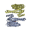 6byjC 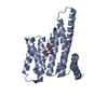 6bykC 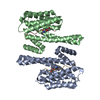 6bzdC  5usk S: Starting model for refinement C: citing same article ( |
|---|---|
| Similar structure data |

-Links
 PDBj
PDBj


- Assembly
| Deposited unit | 
| ||||||||
|---|---|---|---|---|---|---|---|---|---|
| 1 | 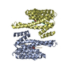
| ||||||||
| 2 | 
| ||||||||
| 3 | 
| ||||||||
| Unit cell |
|
-Components
| #1: Protein | Mass: 27676.920 Da / Num. of mol.: 6 Source method: isolated from a genetically manipulated source Source: (gene. exp.) Homo sapiens (human) / Gene: YWHAG / Production host:  #2: Protein/peptide | Mass: 2015.132 Da / Num. of mol.: 3 / Source method: obtained synthetically / Source: (synth.) synthetic construct (others) #3: Sugar |   Type: D-saccharide, beta linking / Mass: 221.208 Da / Num. of mol.: 3 / Source method: obtained synthetically / Formula: C8H15NO6 Type: D-saccharide, beta linking / Mass: 221.208 Da / Num. of mol.: 3 / Source method: obtained synthetically / Formula: C8H15NO6Has protein modification | Y | |
|---|
-Experimental details
-Experiment
| Experiment | Method: X-RAY DIFFRACTION / Number of used crystals: 1 |
|---|
- Sample preparation
| Crystal | Density Matthews: 3.32 Å3/Da / Density % sol: 62.93 % |
|---|---|
| Crystal grow | Temperature: 298 K / Method: vapor diffusion, hanging drop Details: 29% PEG 4000, 200 mM sodium acetate, 0.1 M Tris pH 8.5 |
-Data collection
| Diffraction | Mean temperature: 100 K |
|---|---|
| Diffraction source | Source: SYNCHROTRON / Site: ALS  / Beamline: 8.3.1 / Wavelength: 1 Å / Beamline: 8.3.1 / Wavelength: 1 Å |
| Detector | Type: ADSC QUANTUM 210r / Detector: CCD / Date: Mar 26, 2017 |
| Radiation | Protocol: SINGLE WAVELENGTH / Monochromatic (M) / Laue (L): M / Scattering type: x-ray |
| Radiation wavelength | Wavelength: 1 Å / Relative weight: 1 |
| Reflection | Resolution: 3.35→112.923 Å / Num. obs: 32119 / % possible obs: 94.5 % / Observed criterion σ(F): 0 / Observed criterion σ(I): 0 / Redundancy: 2.3 % / Biso Wilson estimate: 97.7 Å2 / CC1/2: 0.997 / Rpim(I) all: 0.068 / Rrim(I) all: 0.115 / Rsym value: 0.091 / Net I/σ(I): 6.2 |
| Reflection shell | Resolution: 3.35→3.53 Å / Redundancy: 2.2 % / Mean I/σ(I) obs: 1.2 / Num. unique obs: 4440 / CC1/2: 0.53 / Rpim(I) all: 0.613 / Rrim(I) all: 1.003 / Rsym value: 0.785 / % possible all: 91.5 |
-Phasing
| Phasing | Method: molecular replacement |
|---|
- Processing
| Software |
| |||||||||||||||||||||||||||||||||||||||||||||||||||||||||||||||||||||||||||||||||||||||||||||||||||||||||
|---|---|---|---|---|---|---|---|---|---|---|---|---|---|---|---|---|---|---|---|---|---|---|---|---|---|---|---|---|---|---|---|---|---|---|---|---|---|---|---|---|---|---|---|---|---|---|---|---|---|---|---|---|---|---|---|---|---|---|---|---|---|---|---|---|---|---|---|---|---|---|---|---|---|---|---|---|---|---|---|---|---|---|---|---|---|---|---|---|---|---|---|---|---|---|---|---|---|---|---|---|---|---|---|---|---|---|
| Refinement | Method to determine structure: MOLECULAR REPLACEMENT Starting model: 5USK 5usk Resolution: 3.35→112.923 Å / SU ML: 0.55 / Cross valid method: FREE R-VALUE / σ(F): 1.35 / Phase error: 30.22 / Stereochemistry target values: ML
| |||||||||||||||||||||||||||||||||||||||||||||||||||||||||||||||||||||||||||||||||||||||||||||||||||||||||
| Solvent computation | Shrinkage radii: 0.9 Å / VDW probe radii: 1.11 Å / Solvent model: FLAT BULK SOLVENT MODEL | |||||||||||||||||||||||||||||||||||||||||||||||||||||||||||||||||||||||||||||||||||||||||||||||||||||||||
| Refinement step | Cycle: LAST / Resolution: 3.35→112.923 Å
| |||||||||||||||||||||||||||||||||||||||||||||||||||||||||||||||||||||||||||||||||||||||||||||||||||||||||
| Refine LS restraints |
| |||||||||||||||||||||||||||||||||||||||||||||||||||||||||||||||||||||||||||||||||||||||||||||||||||||||||
| LS refinement shell |
| |||||||||||||||||||||||||||||||||||||||||||||||||||||||||||||||||||||||||||||||||||||||||||||||||||||||||
| Refinement TLS params. | Method: refined / Origin x: 48.6304 Å / Origin y: -14.4459 Å / Origin z: 15.435 Å
| |||||||||||||||||||||||||||||||||||||||||||||||||||||||||||||||||||||||||||||||||||||||||||||||||||||||||
| Refinement TLS group | Selection details: all |
