 Movie
Movie Controller

[English] 日本語
 Yorodumi
- PDB-2cfv: Crystal structure of human protein tyrosine phosphatase receptor ... -
+ Open data

- Basic information
| Entry | Database: PDB / ID: 2cfv | ||||||
|---|---|---|---|---|---|---|---|
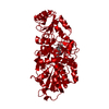
| Title | Crystal structure of human protein tyrosine phosphatase receptor type J | ||||||
 Components Components | HUMAN PROTEIN TYROSINE PHOSPHATASE RECEPTOR TYPE J | ||||||
 Keywords Keywords | HYDROLASE / RECEPTOR TYPE TYROSINE PHOSPHATASE J / PTPRJ / GLYCOPROTEIN / PROTEIN PHOSPHATASE | ||||||
| Function / homology |  Function and homology information Function and homology informationpositive regulation of Fc receptor mediated stimulatory signaling pathway / contact inhibition / gamma-catenin binding / delta-catenin binding / positive regulation of platelet activation / platelet-derived growth factor receptor binding / mitogen-activated protein kinase binding / negative regulation of vascular permeability / platelet formation / negative regulation of MAP kinase activity ...positive regulation of Fc receptor mediated stimulatory signaling pathway / contact inhibition / gamma-catenin binding / delta-catenin binding / positive regulation of platelet activation / platelet-derived growth factor receptor binding / mitogen-activated protein kinase binding / negative regulation of vascular permeability / platelet formation / negative regulation of MAP kinase activity / positive chemotaxis / Phosphorylation of CD3 and TCR zeta chains / negative regulation of platelet-derived growth factor receptor signaling pathway / platelet-derived growth factor receptor signaling pathway / positive regulation of macrophage chemotaxis / peptidyl-tyrosine dephosphorylation / phosphatase activity / oligodendrocyte differentiation / negative regulation of epidermal growth factor receptor signaling pathway / immunological synapse / positive regulation of focal adhesion assembly / negative regulation of T cell receptor signaling pathway / vasculogenesis / regulation of cell adhesion / specific granule membrane / protein-tyrosine-phosphatase / negative regulation of insulin receptor signaling pathway / protein tyrosine phosphatase activity / positive regulation of calcium-mediated signaling / positive regulation of cell adhesion / positive regulation of phagocytosis / Negative regulation of FLT3 / B cell differentiation / axon guidance / negative regulation of cell migration / negative regulation of phosphatidylinositol 3-kinase/protein kinase B signal transduction / negative regulation of cell growth / Negative regulation of MET activity / beta-catenin binding / ruffle membrane / cytokine-mediated signaling pathway / blood coagulation / positive regulation of tumor necrosis factor production / cell-cell junction / cell junction / T cell receptor signaling pathway / glucose homeostasis / heart development / angiogenesis / positive regulation of MAPK cascade / positive regulation of phosphatidylinositol 3-kinase/protein kinase B signal transduction / nuclear body / cadherin binding / negative regulation of cell population proliferation / Neutrophil degranulation / protein kinase binding / nucleolus / cell surface / signal transduction / extracellular exosome / nucleoplasm / plasma membrane Similarity search - Function | ||||||
| Biological species |  HOMO SAPIENS (human) HOMO SAPIENS (human) | ||||||
| Method |  X-RAY DIFFRACTION / SYNCHROTRON / MOLECULAR REPLACEMENT / Resolution: 2.5 Å X-RAY DIFFRACTION / SYNCHROTRON / MOLECULAR REPLACEMENT / Resolution: 2.5 Å | ||||||
 Authors Authors | Debreczeni, J.E. / Barr, A.J. / Eswaran, J. / Ugochukwu, E. / Sundstrom, M. / Weigelt, J. / Arrowsmith, C. / Edwards, A. / Knapp, S. | ||||||
 Citation Citation | Journal: Cell(Cambridge,Mass.) / Year: 2009 Title: Large-Scale Structural Analysis of the Classical Human Protein Tyrosine Phosphatome. Authors: Barr, A.J. / Ugochukwu, E. / Lee, W.H. / King, O.N.F. / Filippakopoulos, P. / Alfano, I. / Savitsky, P. / Burgess-Brown, N.A. / Muller, S. / Knapp, S. | ||||||
| History |
|
- Structure visualization
| Structure viewer | Molecule: MolmilJmol/JSmol |
|---|
- Downloads & links
-Download
| PDBx/mmCIF format | 2cfv.cif.gz | 70.3 KB | Display | PDBx/mmCIF format |
|---|---|---|---|---|
| PDB format | pdb2cfv.ent.gz | 50 KB | Display | PDB format |
| PDBx/mmJSON format | 2cfv.json.gz | Tree view | PDBx/mmJSON format | |
| Others |  Other downloads Other downloads |
-Validation report
| Arichive directory | https://data.pdbj.org/pub/pdb/validation_reports/cf/2cfvftp://data.pdbj.org/pub/pdb/validation_reports/cf/2cfv | HTTPS FTP |
|---|
-Related structure data
| Related structure data |  2ahsSC  2b49C 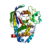 2cjzC  2gjtC  2h4vC  2i75C  2jjdC  2nlkC  2nz6C  2oc3C  2ooqC  2p6xC  2pa5C  2qepC  3b7oC S: Starting model for refinement C: citing same article ( |
|---|---|
| Similar structure data |


-Links
 PDBj
PDBj


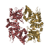
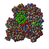
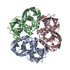
- Assembly
| Deposited unit | 
| ||||||||
|---|---|---|---|---|---|---|---|---|---|
| 1 | 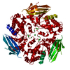
| ||||||||
| Unit cell |
| ||||||||
| Components on special symmetry positions |
|
-Components
| #1: Protein | Mass: 36767.484 Da / Num. of mol.: 1 / Fragment: RESIDUES 1019-1311 Source method: isolated from a genetically manipulated source Source: (gene. exp.) HOMO SAPIENS (human) / Production host:  | ||||
|---|---|---|---|---|---|
| #2: Chemical | ChemComp-NI /   Mass: 58.693 Da / Num. of mol.: 4 / Source method: obtained synthetically / Formula: Ni Mass: 58.693 Da / Num. of mol.: 4 / Source method: obtained synthetically / Formula: Ni#3: Chemical | ChemComp-CL / |   Mass: 35.453 Da / Num. of mol.: 1 / Source method: obtained synthetically / Formula: Cl Mass: 35.453 Da / Num. of mol.: 1 / Source method: obtained synthetically / Formula: Cl#4: Water | ChemComp-HOH / |  Mass: 18.015 Da / Num. of mol.: 25 / Source method: isolated from a natural source / Formula: H2O Mass: 18.015 Da / Num. of mol.: 25 / Source method: isolated from a natural source / Formula: H2O |
-Experimental details
-Experiment
| Experiment | Method: X-RAY DIFFRACTION / Number of used crystals: 1 |
|---|
- Sample preparation
| Crystal | Density Matthews: 2.16 Å3/Da / Density % sol: 43.1 % |
|---|---|
| Crystal grow | Method: vapor diffusion, sitting drop Details: 0.01M NICL2,0.1M TRIS PH 8.5, 1M LI2SO4,150 UL SITTING DROPS |
-Data collection
| Diffraction | Mean temperature: 100 K |
|---|---|
| Diffraction source | Source: SYNCHROTRON / Site: SLS  / Beamline: X10SA / Wavelength: 0.979 / Beamline: X10SA / Wavelength: 0.979 |
| Detector | Type: MARRESEARCH / Detector: CCD / Date: Feb 9, 2006 / Details: MIRROS |
| Radiation | Monochromator: SI / Protocol: SINGLE WAVELENGTH / Monochromatic (M) / Laue (L): M / Scattering type: x-ray |
| Radiation wavelength | Wavelength: 0.979 Å / Relative weight: 1 |
| Reflection | Resolution: 2.5→46.6 Å / Num. obs: 11491 / % possible obs: 98.8 % / Observed criterion σ(I): 3 / Redundancy: 3.76 % / Rmerge(I) obs: 0.06 / Net I/σ(I): 15.25 |
| Reflection shell | Resolution: 2.5→2.6 Å / Redundancy: 3.13 % / Rmerge(I) obs: 0.27 / Mean I/σ(I) obs: 4.18 / % possible all: 91.2 |
- Processing
| Software |
| ||||||||||||||||||||||||||||||||||||||||||||||||||||||||||||||||||||||||||||||||||||||||||||||||||||||||||||||||||||||||||||||||||||||||||||||||||||||||||||||||||||||||||||||||||||||
|---|---|---|---|---|---|---|---|---|---|---|---|---|---|---|---|---|---|---|---|---|---|---|---|---|---|---|---|---|---|---|---|---|---|---|---|---|---|---|---|---|---|---|---|---|---|---|---|---|---|---|---|---|---|---|---|---|---|---|---|---|---|---|---|---|---|---|---|---|---|---|---|---|---|---|---|---|---|---|---|---|---|---|---|---|---|---|---|---|---|---|---|---|---|---|---|---|---|---|---|---|---|---|---|---|---|---|---|---|---|---|---|---|---|---|---|---|---|---|---|---|---|---|---|---|---|---|---|---|---|---|---|---|---|---|---|---|---|---|---|---|---|---|---|---|---|---|---|---|---|---|---|---|---|---|---|---|---|---|---|---|---|---|---|---|---|---|---|---|---|---|---|---|---|---|---|---|---|---|---|---|---|---|---|
| Refinement | Method to determine structure: MOLECULAR REPLACEMENT Starting model: PDB ENTRY 2AHS Resolution: 2.5→63.25 Å / Cor.coef. Fo:Fc: 0.94 / Cor.coef. Fo:Fc free: 0.896 / SU B: 18.803 / SU ML: 0.221 / TLS residual ADP flag: LIKELY RESIDUAL / Cross valid method: THROUGHOUT / ESU R: 0.513 / ESU R Free: 0.285 / Stereochemistry target values: MAXIMUM LIKELIHOOD / Details: HYDROGENS HAVE BEEN ADDED IN THE RIDING POSITIONS.
| ||||||||||||||||||||||||||||||||||||||||||||||||||||||||||||||||||||||||||||||||||||||||||||||||||||||||||||||||||||||||||||||||||||||||||||||||||||||||||||||||||||||||||||||||||||||
| Solvent computation | Ion probe radii: 0.8 Å / Shrinkage radii: 0.8 Å / VDW probe radii: 1.4 Å / Solvent model: MASK | ||||||||||||||||||||||||||||||||||||||||||||||||||||||||||||||||||||||||||||||||||||||||||||||||||||||||||||||||||||||||||||||||||||||||||||||||||||||||||||||||||||||||||||||||||||||
| Displacement parameters | Biso mean: 29.51 Å2
| ||||||||||||||||||||||||||||||||||||||||||||||||||||||||||||||||||||||||||||||||||||||||||||||||||||||||||||||||||||||||||||||||||||||||||||||||||||||||||||||||||||||||||||||||||||||
| Refinement step | Cycle: LAST / Resolution: 2.5→63.25 Å
| ||||||||||||||||||||||||||||||||||||||||||||||||||||||||||||||||||||||||||||||||||||||||||||||||||||||||||||||||||||||||||||||||||||||||||||||||||||||||||||||||||||||||||||||||||||||
| Refine LS restraints |
|