 Movie
Movie Controller

[English] 日本語
 Yorodumi
- PDB-1fpr: CRYSTAL STRUCTURE OF THE COMPLEX FORMED BETWEEN THE CATALYTIC DOM... -
+ Open data

- Basic information
| Entry | Database: PDB / ID: 1fpr | ||||||
|---|---|---|---|---|---|---|---|
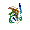
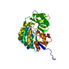
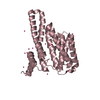
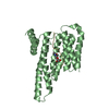
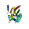
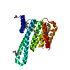
| Title | CRYSTAL STRUCTURE OF THE COMPLEX FORMED BETWEEN THE CATALYTIC DOMAIN OF SHP-1 AND AN IN VITRO PEPTIDE SUBSTRATE PY469 DERIVED FROM SHPS-1. | ||||||
 Components Components |
| ||||||
 Keywords Keywords | SIGNALING PROTEIN / protein tyrosine phosphatase / substrate specificity / residue shift | ||||||
| Function / homology |  Function and homology information Function and homology informationepididymis development / negative regulation of humoral immune response mediated by circulating immunoglobulin / negative regulation of mast cell activation involved in immune response / negative regulation of B cell receptor signaling pathway / regulation of B cell differentiation / CD27 signaling pathway / transmembrane receptor protein tyrosine phosphatase activity / negative regulation of inflammatory response to wounding / phosphorylation-dependent protein binding / negative regulation of lipopolysaccharide-mediated signaling pathway ...epididymis development / negative regulation of humoral immune response mediated by circulating immunoglobulin / negative regulation of mast cell activation involved in immune response / negative regulation of B cell receptor signaling pathway / regulation of B cell differentiation / CD27 signaling pathway / transmembrane receptor protein tyrosine phosphatase activity / negative regulation of inflammatory response to wounding / phosphorylation-dependent protein binding / negative regulation of lipopolysaccharide-mediated signaling pathway / Co-inhibition by BTLA / negative regulation of neutrophil activation / CD22 mediated BCR regulation / Interleukin-37 signaling / regulation of release of sequestered calcium ion into cytosol / positive regulation of cell adhesion mediated by integrin / alpha-beta T cell receptor complex / Signal regulatory protein family interactions / platelet formation / Regulation of KIT signaling / protein dephosphorylation / megakaryocyte development / Signaling by ALK / natural killer cell mediated cytotoxicity / Platelet sensitization by LDL / regulation of type I interferon-mediated signaling pathway / PECAM1 interactions / peptidyl-tyrosine dephosphorylation / non-membrane spanning protein tyrosine phosphatase activity / negative regulation of interleukin-6 production / Regulation of IFNA/IFNB signaling / regulation of G1/S transition of mitotic cell cycle / Interleukin-3, Interleukin-5 and GM-CSF signaling / Co-inhibition by PD-1 / negative regulation of tumor necrosis factor production / T cell proliferation / Interleukin receptor SHC signaling / Regulation of IFNG signaling / negative regulation of T cell receptor signaling pathway / Growth hormone receptor signaling / hematopoietic progenitor cell differentiation / regulation of ERK1 and ERK2 cascade / negative regulation of T cell proliferation / GPVI-mediated activation cascade / T cell costimulation / negative regulation of MAPK cascade / phosphotyrosine residue binding / peptidyl-tyrosine phosphorylation / protein-tyrosine-phosphatase / Nuclear events stimulated by ALK signaling in cancer / protein tyrosine phosphatase activity / cell adhesion molecule binding / negative regulation of innate immune response / negative regulation of angiogenesis / SH2 domain binding / Antigen activates B Cell Receptor (BCR) leading to generation of second messengers / T cell activation / B cell receptor signaling pathway / SH3 domain binding / platelet aggregation / specific granule lumen / Interferon gamma signaling / cytokine-mediated signaling pathway / cell-cell junction / Interferon alpha/beta signaling / tertiary granule lumen / MAPK cascade / mitotic cell cycle / T cell receptor signaling pathway / regulation of apoptotic process / cell differentiation / positive regulation of phosphatidylinositol 3-kinase/protein kinase B signal transduction / G protein-coupled receptor signaling pathway / negative regulation of cell population proliferation / positive regulation of cell population proliferation / Neutrophil degranulation / protein kinase binding / SARS-CoV-2 activates/modulates innate and adaptive immune responses / nucleolus / protein-containing complex / extracellular exosome / extracellular region / nucleoplasm / membrane / nucleus / plasma membrane / cytoplasm / cytosol Similarity search - Function | ||||||
| Biological species |  Homo sapiens (human) Homo sapiens (human) | ||||||
| Method |  X-RAY DIFFRACTION / Resolution: 2.5 Å X-RAY DIFFRACTION / Resolution: 2.5 Å | ||||||
 Authors Authors | Yang, J. / Cheng, Z. / Niu, Z. / Zhao, Z.J. / Zhou, G.W. | ||||||
 Citation Citation | Journal: J.Biol.Chem. / Year: 2000 Title: Structural basis for substrate specificity of protein-tyrosine phosphatase SHP-1. Authors: Yang, J. / Cheng, Z. / Niu, T. / Liang, X. / Zhao, Z.J. / Zhou, G.W. | ||||||
| History |
|
- Structure visualization
| Structure viewer | Molecule: MolmilJmol/JSmol |
|---|
- Downloads & links
-Download
| PDBx/mmCIF format | 1fpr.cif.gz | 71 KB | Display | PDBx/mmCIF format |
|---|---|---|---|---|
| PDB format | pdb1fpr.ent.gz | 52 KB | Display | PDB format |
| PDBx/mmJSON format | 1fpr.json.gz | Tree view | PDBx/mmJSON format | |
| Others |  Other downloads Other downloads |
-Validation report
| Arichive directory | https://data.pdbj.org/pub/pdb/validation_reports/fp/1fprftp://data.pdbj.org/pub/pdb/validation_reports/fp/1fpr | HTTPS FTP |
|---|
-Related structure data
| Similar structure data |
|---|

-Links
 PDBj
PDBj


- Assembly
| Deposited unit | 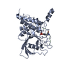
| ||||||||
|---|---|---|---|---|---|---|---|---|---|
| 1 |
| ||||||||
| 2 | 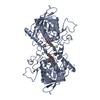
| ||||||||
| Unit cell |
|
-Components
| #1: Protein | Mass: 32558.781 Da / Num. of mol.: 1 / Fragment: CATALYTIC DOMAIN / Mutation: C455S Source method: isolated from a genetically manipulated source Source: (gene. exp.) Homo sapiens (human) / Production host:  |
|---|---|
| #2: Protein/peptide | Mass: 1235.146 Da / Num. of mol.: 1 / Source method: obtained synthetically Details: This peptide was chemically synthesized. The sequence of this peptide occurs naturally in humans (Homo sapiens) |
| Has protein modification | Y |
-Experimental details
-Experiment
| Experiment | Method: X-RAY DIFFRACTION / Number of used crystals: 1 |
|---|
- Sample preparation
| Crystal | Density Matthews: 2.1 Å3/Da / Density % sol: 41.4 % | ||||||||||||||||||||
|---|---|---|---|---|---|---|---|---|---|---|---|---|---|---|---|---|---|---|---|---|---|
| Crystal grow | Temperature: 277 K / Method: vapor diffusion / pH: 8.5 Details: ammonium sulfate, pH 8.5, VAPOR DIFFUSION, temperature 277K | ||||||||||||||||||||
| Crystal grow | *PLUS | ||||||||||||||||||||
| Components of the solutions | *PLUS
|
-Data collection
| Diffraction | Mean temperature: 85 K |
|---|---|
| Diffraction source | Source: ROTATING ANODE / Type: RIGAKU RU300 / Wavelength: 1.5418 |
| Detector | Type: MARRESEARCH / Detector: IMAGE PLATE / Date: Aug 8, 1998 |
| Radiation | Protocol: SINGLE WAVELENGTH / Monochromatic (M) / Laue (L): M / Scattering type: x-ray |
| Radiation wavelength | Wavelength: 1.5418 Å / Relative weight: 1 |
| Reflection | Highest resolution: 2.5 Å / % possible obs: 85 % / Rmerge(I) obs: 0.097 |
| Reflection | *PLUS Num. obs: 8998 / % possible obs: 85 % / Num. measured all: 27345 |
- Processing
| Software |
| ||||||||||||||||||||
|---|---|---|---|---|---|---|---|---|---|---|---|---|---|---|---|---|---|---|---|---|---|
| Refinement | Resolution: 2.5→6 Å / σ(F): 2
| ||||||||||||||||||||
| Refinement step | Cycle: LAST / Resolution: 2.5→6 Å
| ||||||||||||||||||||
| Refine LS restraints |
| ||||||||||||||||||||
| Software | *PLUS Name: X-PLOR / Version: 3.851 / Classification: refinement | ||||||||||||||||||||
| Refinement | *PLUS Highest resolution: 2.5 Å / Lowest resolution: 6 Å / σ(F): 2 / Rfactor all: 0.199 / Rfactor obs: 0.194 | ||||||||||||||||||||
| Solvent computation | *PLUS | ||||||||||||||||||||
| Displacement parameters | *PLUS | ||||||||||||||||||||
| Refine LS restraints | *PLUS Type: x_angle_deg / Dev ideal: 1.4 |
