 Movie
Movie Controller


+ Open data

- Basic information
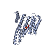
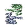
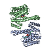
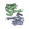
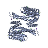
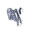
| Entry | Database: PDB / ID: 5oeg | ||||||||||||
|---|---|---|---|---|---|---|---|---|---|---|---|---|---|
| Title | Molecular tweezers modulate 14-3-3 protein-protein interactions | ||||||||||||
 Components Components | 14-3-3 protein sigma | ||||||||||||
 Keywords Keywords | SIGNALING PROTEIN / 14-3-3 / Inhibition / Supramolecular ligand | ||||||||||||
| Function / homology |  Function and homology information Function and homology informationpositive regulation of epidermal cell differentiation / keratinocyte development / regulation of epidermal cell division / protein kinase C inhibitor activity / keratinization / regulation of cell-cell adhesion / keratinocyte proliferation / establishment of skin barrier / Regulation of localization of FOXO transcription factors / negative regulation of keratinocyte proliferation ...positive regulation of epidermal cell differentiation / keratinocyte development / regulation of epidermal cell division / protein kinase C inhibitor activity / keratinization / regulation of cell-cell adhesion / keratinocyte proliferation / establishment of skin barrier / Regulation of localization of FOXO transcription factors / negative regulation of keratinocyte proliferation / phosphoserine residue binding / Activation of BAD and translocation to mitochondria / cAMP/PKA signal transduction / negative regulation of stem cell proliferation / negative regulation of protein localization to plasma membrane / SARS-CoV-2 targets host intracellular signalling and regulatory pathways / negative regulation of protein kinase activity / Chk1/Chk2(Cds1) mediated inactivation of Cyclin B:Cdk1 complex / SARS-CoV-1 targets host intracellular signalling and regulatory pathways / RHO GTPases activate PKNs / positive regulation of protein localization / stem cell proliferation / protein export from nucleus / release of cytochrome c from mitochondria / TP53 Regulates Transcription of Genes Involved in G2 Cell Cycle Arrest / negative regulation of innate immune response / positive regulation of cell adhesion / positive regulation of protein export from nucleus / TP53 Regulates Metabolic Genes / Translocation of SLC2A4 (GLUT4) to the plasma membrane / protein sequestering activity / intrinsic apoptotic signaling pathway in response to DNA damage / intracellular protein localization / regulation of protein localization / sperm midpiece / positive regulation of cell growth / regulation of cell cycle / cadherin binding / protein kinase binding / negative regulation of transcription by RNA polymerase II / signal transduction / : / extracellular exosome / identical protein binding / nucleus / cytosol / cytoplasm Similarity search - Function | ||||||||||||
| Biological species |  Homo sapiens (human) Homo sapiens (human) | ||||||||||||
| Method |  X-RAY DIFFRACTION / MOLECULAR REPLACEMENT / molecular replacement / Resolution: 3.15 Å X-RAY DIFFRACTION / MOLECULAR REPLACEMENT / molecular replacement / Resolution: 3.15 Å | ||||||||||||
 Authors Authors | Bier, D. / Ottmann, C. | ||||||||||||
 Citation Citation | Journal: Nat Chem / Year: 2013 Title: Molecular tweezers modulate 14-3-3 protein-protein interactions. Authors: Bier, D. / Rose, R. / Bravo-Rodriguez, K. / Bartel, M. / Ramirez-Anguita, J.M. / Dutt, S. / Wilch, C. / Klarner, F.G. / Sanchez-Garcia, E. / Schrader, T. / Ottmann, C. | ||||||||||||
| History |
|
- Structure visualization
| Structure viewer | Molecule: MolmilJmol/JSmol |
|---|
- Downloads & links
-Download
| PDBx/mmCIF format | 5oeg.cif.gz | 64.4 KB | Display | PDBx/mmCIF format |
|---|---|---|---|---|
| PDB format | pdb5oeg.ent.gz | 45.8 KB | Display | PDB format |
| PDBx/mmJSON format | 5oeg.json.gz | Tree view | PDBx/mmJSON format | |
| Others |  Other downloads Other downloads |
-Validation report
| Arichive directory | https://data.pdbj.org/pub/pdb/validation_reports/oe/5oegftp://data.pdbj.org/pub/pdb/validation_reports/oe/5oeg | HTTPS FTP |
|---|
-Related structure data
| Related structure data | 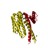 5oehC 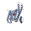 3lw1S S: Starting model for refinement C: citing same article ( |
|---|---|
| Similar structure data |

-Links
 PDBj
PDBj


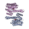
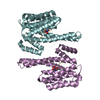
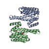
- Assembly
| Deposited unit | 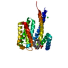
| |||||||||
|---|---|---|---|---|---|---|---|---|---|---|
| 1 | 
| |||||||||
| Unit cell |
| |||||||||
| Components on special symmetry positions |
|
-Components
| #1: Protein | Mass: 26485.865 Da / Num. of mol.: 1 Source method: isolated from a genetically manipulated source Details: AMGS - cleavage artifact / Source: (gene. exp.) Homo sapiens (human) / Gene: SFN, HME1 / Production host:  |
|---|---|
| #2: Chemical | ChemComp-9SZ / (  Mass: 726.646 Da / Num. of mol.: 1 / Source method: obtained synthetically / Formula: C42H32O8P2 Mass: 726.646 Da / Num. of mol.: 1 / Source method: obtained synthetically / Formula: C42H32O8P2 |
| #3: Chemical | ChemComp-CL /   Mass: 35.453 Da / Num. of mol.: 1 / Source method: obtained synthetically / Formula: Cl Mass: 35.453 Da / Num. of mol.: 1 / Source method: obtained synthetically / Formula: Cl |
| #4: Water | ChemComp-HOH /  Mass: 18.015 Da / Num. of mol.: 130 / Source method: isolated from a natural source / Formula: H2O Mass: 18.015 Da / Num. of mol.: 130 / Source method: isolated from a natural source / Formula: H2O |
| Has protein modification | N |
-Experimental details
-Experiment
| Experiment | Method: X-RAY DIFFRACTION / Number of used crystals: 1 |
|---|
- Sample preparation
| Crystal | Density Matthews: 3.57 Å3/Da / Density % sol: 65.55 % |
|---|---|
| Crystal grow | Temperature: 277.15 K / Method: vapor diffusion / pH: 7 / Details: 0.1 M Tris/HCl, 10% PEG 8000, 500 mM MgCl2, pH 7 |
-Data collection
| Diffraction | Mean temperature: 100 K | |||||||||||||||||||||||||||||||||||||||||||||||||
|---|---|---|---|---|---|---|---|---|---|---|---|---|---|---|---|---|---|---|---|---|---|---|---|---|---|---|---|---|---|---|---|---|---|---|---|---|---|---|---|---|---|---|---|---|---|---|---|---|---|---|
| Diffraction source | Source: ROTATING ANODE / Type: RIGAKU MICROMAX-007 / Wavelength: 1.54 Å | |||||||||||||||||||||||||||||||||||||||||||||||||
| Detector | Type: RIGAKU / Detector: IMAGE PLATE / Date: Mar 15, 2010 | |||||||||||||||||||||||||||||||||||||||||||||||||
| Radiation | Protocol: SINGLE WAVELENGTH / Monochromatic (M) / Laue (L): M / Scattering type: x-ray | |||||||||||||||||||||||||||||||||||||||||||||||||
| Radiation wavelength | Wavelength: 1.54 Å / Relative weight: 1 | |||||||||||||||||||||||||||||||||||||||||||||||||
| Reflection | Resolution: 3.15→19.58 Å / Num. obs: 6825 / % possible obs: 99.3 % / Observed criterion σ(I): -3 / Redundancy: 3.584 % / Biso Wilson estimate: 30.196 Å2 / Rmerge(I) obs: 0.086 / Rrim(I) all: 0.102 / Χ2: 0.926 / Net I/σ(I): 16.05 | |||||||||||||||||||||||||||||||||||||||||||||||||
| Reflection shell | Diffraction-ID: 1
|
-Phasing
| Phasing | Method: molecular replacement | |||||||||
|---|---|---|---|---|---|---|---|---|---|---|
| Phasing MR | Model details: Phaser MODE: MR_AUTO
|
- Processing
| Software |
| ||||||||||||||||||||||||||||||||||||||||||||||||||||||||||||
|---|---|---|---|---|---|---|---|---|---|---|---|---|---|---|---|---|---|---|---|---|---|---|---|---|---|---|---|---|---|---|---|---|---|---|---|---|---|---|---|---|---|---|---|---|---|---|---|---|---|---|---|---|---|---|---|---|---|---|---|---|---|
| Refinement | Method to determine structure: MOLECULAR REPLACEMENT Starting model: 3LW1 Resolution: 3.15→19.58 Å / Cor.coef. Fo:Fc: 0.931 / Cor.coef. Fo:Fc free: 0.844 / SU B: 18.689 / SU ML: 0.321 / Cross valid method: THROUGHOUT / σ(F): 0 / ESU R Free: 0.471 Details: HYDROGENS HAVE BEEN ADDED IN THE RIDING POSITIONS U VALUES : REFINED INDIVIDUALLY
| ||||||||||||||||||||||||||||||||||||||||||||||||||||||||||||
| Solvent computation | Ion probe radii: 0.8 Å / Shrinkage radii: 0.8 Å / VDW probe radii: 1.2 Å | ||||||||||||||||||||||||||||||||||||||||||||||||||||||||||||
| Displacement parameters | Biso max: 88.25 Å2 / Biso mean: 39.985 Å2 / Biso min: 1 Å2
| ||||||||||||||||||||||||||||||||||||||||||||||||||||||||||||
| Refinement step | Cycle: final / Resolution: 3.15→19.58 Å
| ||||||||||||||||||||||||||||||||||||||||||||||||||||||||||||
| Refine LS restraints |
| ||||||||||||||||||||||||||||||||||||||||||||||||||||||||||||
| LS refinement shell | Resolution: 3.15→3.23 Å / Rfactor Rfree error: 0 / Total num. of bins used: 20
|
