 Movie
Movie Controller


+ Open data

- Basic information
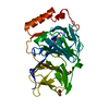
| Entry | Database: PDB / ID: 1rpm | ||||||
|---|---|---|---|---|---|---|---|
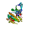
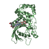
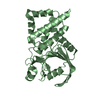
| Title | HUMAN RECEPTOR PROTEIN TYROSINE PHOSPHATASE MU, DOMAIN 1 | ||||||
 Components Components | RECEPTOR PROTEIN TYROSINE PHOSPHATASE MU | ||||||
 Keywords Keywords | RECEPTOR / PHOSPHATASE / SIGNAL TRANSDUCTION / ADHESION / HYDROLASE | ||||||
| Function / homology |  Function and homology information Function and homology informationtransmembrane receptor protein tyrosine phosphatase activity / retina layer formation / positive regulation of D-glucose transmembrane transport / negative regulation of endothelial cell migration / retinal ganglion cell axon guidance / negative regulation of endothelial cell proliferation / homophilic cell-cell adhesion / phosphatase activity / protein-tyrosine-phosphatase / protein tyrosine phosphatase activity ...transmembrane receptor protein tyrosine phosphatase activity / retina layer formation / positive regulation of D-glucose transmembrane transport / negative regulation of endothelial cell migration / retinal ganglion cell axon guidance / negative regulation of endothelial cell proliferation / homophilic cell-cell adhesion / phosphatase activity / protein-tyrosine-phosphatase / protein tyrosine phosphatase activity / negative regulation of angiogenesis / adherens junction / neuron projection development / cell-cell junction / lamellipodium / cadherin binding / response to xenobiotic stimulus / perinuclear region of cytoplasm / signal transduction / identical protein binding / plasma membrane / cytoplasm Similarity search - Function | ||||||
| Biological species |  Homo sapiens (human) Homo sapiens (human) | ||||||
| Method |  X-RAY DIFFRACTION / SYNCHROTRON / MOLECULAR REPLACEMENT / Resolution: 2.3 Å X-RAY DIFFRACTION / SYNCHROTRON / MOLECULAR REPLACEMENT / Resolution: 2.3 Å | ||||||
 Authors Authors | Hoffmann, K.M.V. / Tonks, N.K. / Barford, D. | ||||||
 Citation Citation | Journal: J.Biol.Chem. / Year: 1997 Title: The crystal structure of domain 1 of receptor protein-tyrosine phosphatase mu. Authors: Hoffmann, K.M. / Tonks, N.K. / Barford, D. | ||||||
| History |
|
- Structure visualization
| Structure viewer | Molecule: MolmilJmol/JSmol |
|---|
- Downloads & links
-Download
| PDBx/mmCIF format | 1rpm.cif.gz | 128.6 KB | Display | PDBx/mmCIF format |
|---|---|---|---|---|
| PDB format | pdb1rpm.ent.gz | 99.2 KB | Display | PDB format |
| PDBx/mmJSON format | 1rpm.json.gz | Tree view | PDBx/mmJSON format | |
| Others |  Other downloads Other downloads |
-Validation report
| Arichive directory | https://data.pdbj.org/pub/pdb/validation_reports/rp/1rpmftp://data.pdbj.org/pub/pdb/validation_reports/rp/1rpm | HTTPS FTP |
|---|
-Related structure data
| Related structure data | 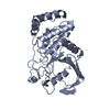 1yfoS S: Starting model for refinement |
|---|---|
| Similar structure data |


-Links
 PDBj
PDBj


- Assembly
| Deposited unit | 
| ||||||||
|---|---|---|---|---|---|---|---|---|---|
| 1 | 
| ||||||||
| 2 | 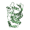
| ||||||||
| Unit cell |
| ||||||||
| Noncrystallographic symmetry (NCS) | NCS oper: (Code: given Matrix: (-0.0988, -0.0011, 0.9951), Vector: |
-Components
| #1: Protein | Mass: 32151.496 Da / Num. of mol.: 2 / Fragment: CYTOSOLIC MEMBRANE PROXIMAL CATALYTIC DOMAIN Source method: isolated from a genetically manipulated source Source: (gene. exp.) Homo sapiens (human) / Production host:  #2: Water | ChemComp-HOH / |  Mass: 18.015 Da / Num. of mol.: 350 / Source method: isolated from a natural source / Formula: H2O Mass: 18.015 Da / Num. of mol.: 350 / Source method: isolated from a natural source / Formula: H2O |
|---|
-Experimental details
-Experiment
| Experiment | Method: X-RAY DIFFRACTION / Number of used crystals: 1 |
|---|
- Sample preparation
| Crystal | Density Matthews: 2.56 Å3/Da / Density % sol: 52 % | ||||||||||||||||||||||||||||||
|---|---|---|---|---|---|---|---|---|---|---|---|---|---|---|---|---|---|---|---|---|---|---|---|---|---|---|---|---|---|---|---|
| Crystal grow | pH: 5.5 Details: PROTEIN WAS CRYSTALLIZED FROM 750 MM SODIUM CITRATE, PH 5.5, 2 MM DTT, 0.5 MM EDTA | ||||||||||||||||||||||||||||||
| Crystal | *PLUS | ||||||||||||||||||||||||||||||
| Crystal grow | *PLUS Temperature: 4 ℃ / Method: unknown | ||||||||||||||||||||||||||||||
| Components of the solutions | *PLUS
|
-Data collection
| Diffraction | Mean temperature: 100 K |
|---|---|
| Diffraction source | Source: SYNCHROTRON / Site: SRS  / Beamline: PX7.2 / Wavelength: 1.488 / Beamline: PX7.2 / Wavelength: 1.488 |
| Detector | Type: MAR scanner 300 mm plate / Detector: IMAGE PLATE / Date: Apr 1, 1997 |
| Radiation | Monochromator: GE(111) / Monochromatic (M) / Laue (L): M / Scattering type: x-ray |
| Radiation wavelength | Wavelength: 1.488 Å / Relative weight: 1 |
| Reflection | Resolution: 2.3→15 Å / Num. obs: 26733 / % possible obs: 90 % / Redundancy: 4 % / Rsym value: 0.029 / Net I/σ(I): 25.2 |
| Reflection shell | Resolution: 2.3→2.38 Å / Rsym value: 0.064 / % possible all: 92 |
| Reflection | *PLUS Lowest resolution: 50 Å / Num. measured all: 112065 / Rmerge(I) obs: 0.03 |
- Processing
| Software |
| ||||||||||||||||||||||||||||||||||||||||||||||||||||||||||||
|---|---|---|---|---|---|---|---|---|---|---|---|---|---|---|---|---|---|---|---|---|---|---|---|---|---|---|---|---|---|---|---|---|---|---|---|---|---|---|---|---|---|---|---|---|---|---|---|---|---|---|---|---|---|---|---|---|---|---|---|---|---|
| Refinement | Method to determine structure: MOLECULAR REPLACEMENT Starting model: PDB ENTRY 1YFO Resolution: 2.3→8 Å / Data cutoff high absF: 100000 / Data cutoff low absF: 0.1 / Cross valid method: FREE R / σ(F): 2
| ||||||||||||||||||||||||||||||||||||||||||||||||||||||||||||
| Refinement step | Cycle: LAST / Resolution: 2.3→8 Å
| ||||||||||||||||||||||||||||||||||||||||||||||||||||||||||||
| Refine LS restraints |
| ||||||||||||||||||||||||||||||||||||||||||||||||||||||||||||
| Refine LS restraints NCS | NCS model details: RESTRAINTS | ||||||||||||||||||||||||||||||||||||||||||||||||||||||||||||
| LS refinement shell | Resolution: 2.3→8 Å / Total num. of bins used: 8 | ||||||||||||||||||||||||||||||||||||||||||||||||||||||||||||
| Xplor file |
|
