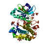
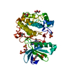
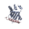
SHEET THE SHEET STRUCTURE OF THIS MOLECULE IS BIFURCATED. IN ORDER TO REPRESENT THIS FEATURE IN ... SHEET THE SHEET STRUCTURE OF THIS MOLECULE IS BIFURCATED. IN ORDER TO REPRESENT THIS FEATURE IN THE SHEET RECORDS BELOW, TWO SHEETS ARE DEFINED.
Protocol: SINGLE WAVELENGTH / Monochromatic (M) / Laue (L): M / Scattering type: x-ray
Radiation wavelength
Wavelength: 1.5418 Å / Relative weight: 1
Reflection
Resolution: 1.45→30 Å / Num. obs: 95063 / % possible obs: 97.5 % / Observed criterion σ(I): 2 / Redundancy: 4.5 % / Rmerge(I) obs: 0.05 / Net I/σ(I): 13.2
Reflection shell
Resolution: 1.45→1.5 Å / Redundancy: 3 % / Rmerge(I) obs: 0.4 / Mean I/σ(I) obs: 2.7 / % possible all: 80.1
-
Processing
Software
Name
Version
Classification
REFMAC
5.2.0003
refinement
CrystalClear
datareduction
d*TREK
datascaling
AMoRE
phasing
Refinement
Method to determine structure: MOLECULAR REPLACEMENT Starting model: PDK1PH INSP4 MODEL Resolution: 1.45→30 Å / Cor.coef. Fo:Fc: 0.976 / Cor.coef. Fo:Fc free: 0.96 / SU B: 3.811 / SU ML: 0.064 / Cross valid method: THROUGHOUT / ESU R: 0.082 / ESU R Free: 0.074 / Stereochemistry target values: MAXIMUM LIKELIHOOD Details: HYDROGENS HAVE BEEN ADDED IN THE RIDING POSITIONS. THE SIDE CHAINS OF SOME DISORDERED RESIDUES WERE REFINED EITHER WITH THE OCCUPANCY SET TO 0.02, OR THE RESIDUE WAS MUTATED TO ALA.
Rfactor
Num. reflection
% reflection
Selection details
Rfree
0.206
934
1 %
RANDOM
Rwork
0.156
-
-
-
obs
0.157
94091
97.4 %
-
Solvent computation
Ion probe radii: 0.8 Å / Shrinkage radii: 0.8 Å / VDW probe radii: 1.2 Å / Solvent model: BABINET MODEL WITH MASK
 Movie
Movie Controller


 Open data
 Basic information
 Components
 Keywords
 Function and homology information
 HOMO SAPIENS (human)
 X-RAY DIFFRACTION /
  Authors
 Citation
 Structure visualization
 Downloads & links
 Other downloads
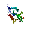


 PDBj
PDBj


 Assembly


 Mass: 92.094 Da / Num. of mol.: 4 / Source method: obtained synthetically / Formula: C3H8O3
 Mass: 96.063 Da / Num. of mol.: 10 / Source method: obtained synthetically / Formula: SO4
 Mass: 18.015 Da / Num. of mol.: 715 / Source method: isolated from a natural source / Formula: H2O
 Sample preparation
 Processing