[日本語] English
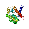
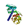
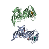
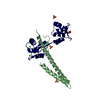
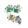
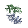
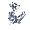
- PDB-1tjt: X-ray structure of the human alpha-actinin isoform 3 at 2.2A reso... -

+
データを開く


IDまたはキーワード:

読み込み中...

-
基本情報

登録情報
データベース: PDB / ID: 1tjt
タイトルX-ray structure of the human alpha-actinin isoform 3 at 2.2A resolution
要素Alpha-actinin 3
キーワードCONTRACTILE PROTEIN / Calponin homology domain / actin binding domain
機能・相同性
機能・相同性情報


positive regulation of glucose catabolic process to lactate via pyruvate / negative regulation of relaxation of muscle / regulation of the force of skeletal muscle contraction / skeletal muscle atrophy / positive regulation of skeletal muscle fiber development / positive regulation of skeletal muscle tissue growth / response to denervation involved in regulation of muscle adaptation / positive regulation of fast-twitch skeletal muscle fiber contraction / transition between fast and slow fiber / positive regulation of bone mineralization involved in bone maturation ...positive regulation of glucose catabolic process to lactate via pyruvate / negative regulation of relaxation of muscle / regulation of the force of skeletal muscle contraction / skeletal muscle atrophy / positive regulation of skeletal muscle fiber development / positive regulation of skeletal muscle tissue growth / response to denervation involved in regulation of muscle adaptation / positive regulation of fast-twitch skeletal muscle fiber contraction / transition between fast and slow fiber / positive regulation of bone mineralization involved in bone maturation / muscle cell development / negative regulation of oxidative phosphorylation / focal adhesion assembly / Striated Muscle Contraction / bone morphogenesis / negative regulation of glycolytic process / Nephrin family interactions / negative regulation of cold-induced thermogenesis / negative regulation of calcineurin-NFAT signaling cascade / structural constituent of muscle / regulation of aerobic respiration / cortical actin cytoskeleton / pseudopodium / brush border / cell projection / actin filament / Z disc / actin filament binding / integrin binding / cell junction / actin cytoskeleton organization / regulation of apoptotic process / transmembrane transporter binding / focal adhesion / calcium ion binding / extracellular exosome / identical protein binding / plasma membrane / cytosol
類似検索 - 分子機能
Calponin-like domain / EF-hand, Ca insensitive / Ca2+ insensitive EF hand / Ca2+ insensitive EF hand / Actin-binding Protein, T-fimbrin; domain 1 / Spectrin repeat / Spectrin repeat / Spectrin/alpha-actinin / Actinin-type actin-binding domain signature 1. / Actinin-type actin-binding domain signature 2. ...Calponin-like domain / EF-hand, Ca insensitive / Ca2+ insensitive EF hand / Ca2+ insensitive EF hand / Actin-binding Protein, T-fimbrin; domain 1 / Spectrin repeat / Spectrin repeat / Spectrin/alpha-actinin / Actinin-type actin-binding domain signature 1. / Actinin-type actin-binding domain signature 2. / Spectrin repeats / Actinin-type actin-binding domain, conserved site / Calponin homology domain / Calponin homology (CH) domain / Calponin homology domain / CH domain superfamily / Calponin homology (CH) domain profile. / EF-hand, calcium binding motif / EF-hand calcium-binding domain profile. / EF-hand domain / EF-hand domain pair / Orthogonal Bundle / Mainly Alpha
類似検索 - ドメイン・相同性
生物種Homo sapiens (ヒト)
手法X線回折 / 分子置換 / 解像度: 2.19 Å
データ登録者Franzot, G. / Sjoblom, B. / Gautel, M. / Djinovic Carugo, K.
引用ジャーナル: J.Mol.Biol. / : 2005
タイトル: The crystal structure of the actin binding domain from alpha-actinin in its closed conformation: structural insight into phospholipid regulation of alpha-actinin
著者: Franzot, G. / Sjoblom, B. / Gautel, M. / Djinovic Carugo, K.
履歴
登録2004年6月7日登録サイト: RCSB / 処理サイト: PDBJ
改定 1.02005年5月17日Provider: repository / タイプ: Initial release
改定 1.12008年4月30日Group: Version format compliance
改定 1.22011年7月13日Group: Version format compliance
改定 1.32017年10月11日Group: Refinement description / カテゴリ: software / Item: _software.classification / _software.name
改定 1.42023年10月25日Group: Data collection / Database references / Refinement description
カテゴリ: chem_comp_atom / chem_comp_bond ...chem_comp_atom / chem_comp_bond / database_2 / pdbx_initial_refinement_model / struct_ref_seq_dif
Item: _database_2.pdbx_DOI / _database_2.pdbx_database_accession / _struct_ref_seq_dif.details

-
構造の表示

構造ビューア分子:
MolmilJmol/JSmol

ダウンロードとリンク

-
集合体

登録構造単位
A: Alpha-actinin 3


分子量 (理論値)分子数
合計 (水以外)28,6211
ポリマ-28,6211
非ポリマー00
3,477193
1
A: Alpha-actinin 3

A: Alpha-actinin 3


分子量 (理論値)分子数
合計 (水以外)57,2422
ポリマ-57,2422
非ポリマー00
362
タイプ名称対称操作
identity operation1_555x,y,z1
crystal symmetry operation2_646-x+1,y-1/2,-z+11
単位格子
Length a, b, c (Å)46.310, 38.540, 65.480
Angle α, β, γ (deg.)90.00, 99.72, 90.00
Int Tables number4
Space group name H-MP1211

-
要素

#1: タンパク質 Alpha-actinin 3 / Alpha actinin skeletal muscle isoform 3 / F-actin cross linking protein


分子量: 28620.836 Da / 分子数: 1 / 断片: actin binding domain / 由来タイプ: 組換発現 / 由来: (組換発現) Homo sapiens (ヒト) / 遺伝子: ACTN3 / 発現宿主: Escherichia coli (大腸菌) / 参照: UniProt: Q08043
#2: 水 ChemComp-HOH / water


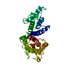
分子量: 18.015 Da / 分子数: 193 / 由来タイプ: 天然 / : H2O

-
実験情報

-
実験

実験手法: X線回折 / 使用した結晶の数: 1

-
試料調製

結晶マシュー密度: 2.01 Å3/Da / 溶媒含有率: 40 %
結晶化温度: 298 K / 手法: 蒸気拡散法, ハンギングドロップ法 / pH: 6.5
詳細: peg 4000, sodium cacodylate, pH 6.5, VAPOR DIFFUSION, HANGING DROP, temperature 298.0K

-
データ収集

回折平均測定温度: 100 K
放射光源由来: 回転陽極 / タイプ: ELLIOTT GX-21 / 波長: 1.5418 Å
検出器タイプ: MARRESEARCH / 検出器: IMAGE PLATE
放射プロトコル: SINGLE WAVELENGTH / 単色(M)・ラウエ(L): M / 散乱光タイプ: x-ray
放射波長波長: 1.5418 Å / 相対比: 1
反射解像度: 2.19→34.61 Å / Num. all: 11674 / Num. obs: 11674 / % possible obs: 97.4 % / Observed criterion σ(F): 0 / Observed criterion σ(I): 0 / 冗長度: 2.8 % / Biso Wilson estimate: 8.6 Å2 / Rmerge(I) obs: 0.089
反射 シェル解像度: 2.19→2.33 Å / 冗長度: 2.8 % / Rmerge(I) obs: 0.186 / % possible all: 93.1

-
解析

ソフトウェア
名称バージョン分類
CNS1.1精密化
MAR345データ収集
XDSデータスケーリング
AMoRE位相決定
精密化構造決定の手法: 分子置換
開始モデル: 1QAG for CH1 domain and 1BKR for CH2 domain
解像度: 2.19→34.61 Å / Rfactor Rfree error: 0.008 / Data cutoff high absF: 867794.42 / Data cutoff low absF: 0 / Isotropic thermal model: RESTRAINED / 交差検証法: THROUGHOUT / σ(F): 0 / 立体化学のターゲット値: Engh & Huber
Rfactor反射数%反射Selection details
Rfree0.273 1197 10.3 %RANDOM
Rwork0.227 ---
obs0.227 11674 97.4 %-
all-11674 --
溶媒の処理溶媒モデル: FLAT MODEL / Bsol: 53.0791 Å2 / ksol: 0.35091 e/Å3
原子変位パラメータBiso mean: 17.8 Å2
Baniso -1Baniso -2Baniso -3
1-0.05 Å20 Å2-2.18 Å2
2--3.22 Å20 Å2
3----3.28 Å2
Refine analyze
FreeObs
Luzzati coordinate error0.34 Å0.26 Å
Luzzati d res low-5 Å
Luzzati sigma a0.29 Å0.19 Å
精密化ステップサイクル: LAST / 解像度: 2.19→34.61 Å
タンパク質核酸リガンド溶媒全体
原子数1799 0 0 193 1992
拘束条件
Refine-IDタイプDev idealDev ideal target
X-RAY DIFFRACTIONc_bond_d0.007
X-RAY DIFFRACTIONc_bond_d_na
X-RAY DIFFRACTIONc_bond_d_prot
X-RAY DIFFRACTIONc_angle_d
X-RAY DIFFRACTIONc_angle_d_na
X-RAY DIFFRACTIONc_angle_d_prot
X-RAY DIFFRACTIONc_angle_deg1.1
X-RAY DIFFRACTIONc_angle_deg_na
X-RAY DIFFRACTIONc_angle_deg_prot
X-RAY DIFFRACTIONc_dihedral_angle_d20.8
X-RAY DIFFRACTIONc_dihedral_angle_d_na
X-RAY DIFFRACTIONc_dihedral_angle_d_prot
X-RAY DIFFRACTIONc_improper_angle_d0.75
X-RAY DIFFRACTIONc_improper_angle_d_na
X-RAY DIFFRACTIONc_improper_angle_d_prot
X-RAY DIFFRACTIONc_mcbond_it1.971.5
X-RAY DIFFRACTIONc_mcangle_it2.372
X-RAY DIFFRACTIONc_scbond_it2.912
X-RAY DIFFRACTIONc_scangle_it3.622.5
LS精密化 シェル解像度: 2.19→2.33 Å / Rfactor Rfree error: 0.022 / Total num. of bins used: 6
Rfactor反射数%反射
Rfree0.305 191 10.3 %
Rwork0.249 1657 -
obs--93.1 %
Xplor file
Refine-IDSerial noParam fileTopol file
X-RAY DIFFRACTION1PROTEIN_REP.PARAMPROTEIN.TOP
X-RAY DIFFRACTION2WATER_REP.PARAM

+
万見について

-
お知らせ

-
2022年2月9日: EMDBエントリの付随情報ファイルのフォーマットが新しくなりました

EMDBエントリの付随情報ファイルのフォーマットが新しくなりました

  • EMDBのヘッダファイルのバージョン3が、公式のフォーマットとなりました。
  • これまでは公式だったバージョン1.9は、アーカイブから削除されます。

関連情報:EMDBヘッダ

外部リンク:wwPDBはEMDBデータモデルのバージョン3へ移行します

-
2020年8月12日: 新型コロナ情報

新型コロナ情報

URL: https://pdbjlvh1.pdbj.org/emnavi/covid19.php

新ページ: EM Navigatorに新型コロナウイルスの特設ページを開設しました。

関連情報:Covid-19情報 / 2020年3月5日: 新型コロナウイルスの構造データ

+
2020年3月5日: 新型コロナウイルスの構造データ

新型コロナウイルスの構造データ

関連情報:万見生物種 / 2020年8月12日: 新型コロナ情報

外部リンク:COVID-19特集ページ - PDBj / 今月の分子2020年2月:コロナウイルスプロテーアーゼ

+
2019年1月31日: EMDBのIDの桁数の変更

EMDBのIDの桁数の変更

  • EMDBエントリに付与されているアクセスコード(EMDB-ID)は4桁の数字(例、EMD-1234)でしたが、間もなく枯渇します。これまでの4桁のID番号は4桁のまま変更されませんが、4桁の数字を使い切った後に発行されるIDは5桁以上の数字(例、EMD-12345)になります。5桁のIDは2019年の春頃から発行される見通しです。
  • EM Navigator/万見では、接頭語「EMD-」は省略されています。

関連情報:Q: 「EMD」とは何ですか? / 万見/EM NavigatorにおけるID/アクセスコードの表記

外部リンク:EMDB Accession Codes are Changing Soon! / PDBjへお問い合わせ

+
2017年7月12日: PDB大規模アップデート

PDB大規模アップデート

  • 新バージョンのPDBx/mmCIF辞書形式に基づくデータがリリースされました。
  • 今回の更新はバージョン番号が4から5になる大規模なもので、全エントリデータの書き換えが行われる「Remediation」というアップデートに該当します。
  • このバージョンアップで、電子顕微鏡の実験手法に関する多くの項目の書式が改定されました(例:em_softwareなど)。
  • EM NavigatorとYorodumiでも、この改定に基づいた表示内容になります。

外部リンク:wwPDB Remediation / OneDepデータ基準に準拠した、より強化された内容のモデル構造ファイルが、PDBアーカイブで公開されました。

-
万見 (Yorodumi)

幾万の構造データを、幾万の視点から

  • 万見(Yorodumi)は、EMDB/PDB/SASBDBなどの構造データを閲覧するためのページです。
  • EM Navigatorの詳細ページの後継、Omokage検索のフロントエンドも兼ねています。

関連情報:EMDB / PDB / SASBDB / 3つのデータバンクの比較 / 万見検索 / 2016年8月31日: 新しいEM Navigatorと万見 / 万見文献 / Jmol/JSmol / 機能・相同性情報 / 新しいEM Navigatorと万見の変更点

他の情報も見る