Entry Database : PDB / ID : 1s4yTitle Crystal structure of the activin/actrIIb extracellular domain Activin receptor type IIB precursor Inhibin beta A chain Keywords / / / / / Function / homology Function Domain/homology Component
/ / / / / / / / / / / / / / / / / / / / / / / / / / / / / / / / / / / / / / / / / / / / / / / / / / / / / / / / / / / / / / / / / / / / / / / / / / / / / / / / / / / / / / / / / / / / / / / / / / / / / / / / / / / / / / / / / / / / / / / / / / / / / / / / / / / / / / / / / / / / / / / Biological species Mus musculus (house mouse)Homo sapiens (human)Method / / / Resolution : 2.3 Å Authors Greenwald, J. / Vega, M.E. / Allendorph, G.P. / Fischer, W.H. / Vale, W. / Choe, S. / Joint Center for Structural Genomics (JCSG) Journal : Mol.Cell / Year : 2004Title : A Flexible Activin Explains the Membrane-Dependent Cooperative Assembly of TGF-beta Family Receptors.Authors : Greenwald, J. / Vega, M.E. / Allendorph, G.P. / Fischer, W.H. / Vale, W. / Choe, S. History Deposition Jan 19, 2004 Deposition site / Processing site Revision 1.0 Aug 10, 2004 Provider / Type Revision 1.1 Apr 29, 2008 Group Revision 1.2 Jul 13, 2011 Group / Version format complianceRevision 1.3 Nov 13, 2024 Group / Database references / Structure summaryCategory chem_comp_atom / chem_comp_bond ... chem_comp_atom / chem_comp_bond / database_2 / pdbx_entry_details / pdbx_modification_feature Item / _database_2.pdbx_database_accession
Show all Show less
 Movie
Movie Controller


 Open data
 Basic information
 Components
 Keywords
 Function and homology information
 Homo sapiens (human)
 X-RAY DIFFRACTION /
  Authors
 Citation
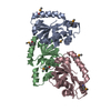
 Structure visualization
 Downloads & links
 Other downloads
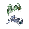


 PDBj
PDBj


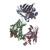
 Assembly

 Cricetulus griseus (Chinese hamster) / References: UniProt: P08476
 Mass: 18.015 Da / Num. of mol.: 177 / Source method: isolated from a natural source / Formula: H2O
 Sample preparation
 / Beamline: 8.2.2 / Wavelength: 1.0332 Å
 Processing