 Movie
Movie Controller

[English] 日本語
 Yorodumi
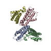
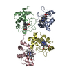
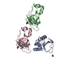
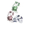
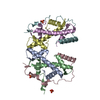
- PDB-1bwu: MANNOSE-SPECIFIC AGGLUTININ (LECTIN) FROM GARLIC (ALLIUM SATIVUM)... -
+ Open data

- Basic information
| Entry | Database: PDB / ID: 1bwu | ||||||
|---|---|---|---|---|---|---|---|
| Title | MANNOSE-SPECIFIC AGGLUTININ (LECTIN) FROM GARLIC (ALLIUM SATIVUM) BULBS COMPLEXED WITH ALPHA-D-MANNOSE | ||||||
 Components Components | (PROTEIN (AGGLUTININ)) x 4 | ||||||
 Keywords Keywords | PLANT PROTEIN / BULB LECTIN / MANNOSE | ||||||
| Function / homology |  Function and homology information Function and homology information | ||||||
| Biological species |  | ||||||
| Method |  X-RAY DIFFRACTION / MOLECULAR REPLACEMENT / Resolution: 2.8 Å X-RAY DIFFRACTION / MOLECULAR REPLACEMENT / Resolution: 2.8 Å | ||||||
 Authors Authors | Chandra, N.R. / Ramachandraiah, G. / Bachhawat, K. / Dam, T.K. / Surolia, A. / Vijayan, M. | ||||||
 Citation Citation | Journal: J.Mol.Biol. / Year: 1999 Title: Crystal structure of a dimeric mannose-specific agglutinin from garlic: quaternary association and carbohydrate specificity. Authors: Chandra, N.R. / Ramachandraiah, G. / Bachhawat, K. / Dam, T.K. / Surolia, A. / Vijayan, M. #1: Journal: Acta Crystallogr.,Sect.D / Year: 1997Title: Crystallization and Preliminary Crystallographic Studies on the Mannose- Specific Lectin from Garlic Authors: Chandra, N.R. / Dam, T.K. / Surolia, A. / Vijayan, M. | ||||||
| History |
|
- Structure visualization
| Structure viewer | Molecule: MolmilJmol/JSmol |
|---|
- Downloads & links
-Download
| PDBx/mmCIF format | 1bwu.cif.gz | 102.7 KB | Display | PDBx/mmCIF format |
|---|---|---|---|---|
| PDB format | pdb1bwu.ent.gz | 80.5 KB | Display | PDB format |
| PDBx/mmJSON format | 1bwu.json.gz | Tree view | PDBx/mmJSON format | |
| Others |  Other downloads Other downloads |
-Validation report
| Arichive directory | https://data.pdbj.org/pub/pdb/validation_reports/bw/1bwuftp://data.pdbj.org/pub/pdb/validation_reports/bw/1bwu | HTTPS FTP |
|---|
-Related structure data
| Related structure data | 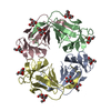 1msaS S: Starting model for refinement |
|---|---|
| Similar structure data |


-Links
 PDBj
PDBj


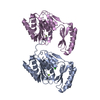
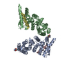
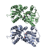
- Assembly
| Deposited unit | 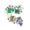
| ||||||||
|---|---|---|---|---|---|---|---|---|---|
| 1 |
| ||||||||
| 2 | 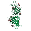
| ||||||||
| 3 | 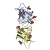
| ||||||||
| Unit cell |
| ||||||||
| Details | SUBUNITS, CHAINS A AND D and CHAINS P AND Q, FORM 2 INDEPENDENT HETERO-DIMERS IN THE ASYMMETRIC UNIT |
-Components
-Protein , 4 types, 4 molecules ADPQ
| #1: Protein | Mass: 11865.071 Da / Num. of mol.: 1 / Source method: isolated from a natural source / Source: (natural) |
|---|---|
| #2: Protein | Mass: 12124.300 Da / Num. of mol.: 1 / Source method: isolated from a natural source / Source: (natural) |
| #3: Protein | Mass: 11678.851 Da / Num. of mol.: 1 / Source method: isolated from a natural source / Source: (natural) |
| #4: Protein | Mass: 12010.171 Da / Num. of mol.: 1 / Source method: isolated from a natural source / Source: (natural) |
-Sugars / Non-polymers , 2 types, 151 molecules 

| #5: Sugar | ChemComp-MAN /  Type: D-saccharide, alpha linking / Mass: 180.156 Da / Num. of mol.: 14 / Source method: obtained synthetically / Formula: C6H12O6 Type: D-saccharide, alpha linking / Mass: 180.156 Da / Num. of mol.: 14 / Source method: obtained synthetically / Formula: C6H12O6#6: Water | ChemComp-HOH / | Mass: 18.015 Da / Num. of mol.: 137 / Source method: isolated from a natural source / Formula: H2O |
|---|
-Details
| Has protein modification | Y |
|---|
-Experimental details
-Experiment
| Experiment | Method: X-RAY DIFFRACTION / Number of used crystals: 1 |
|---|
- Sample preparation
| Crystal | Density Matthews: 3.58 Å3/Da / Density % sol: 65.6 % | |||||||||||||||||||||||||
|---|---|---|---|---|---|---|---|---|---|---|---|---|---|---|---|---|---|---|---|---|---|---|---|---|---|---|
| Crystal grow | Temperature: 298 K / Method: vapor diffusion, hanging drop / pH: 7 Details: 20% PEG8000, 5.5 MG/ML PROTEIN, 10MM MANNOSE, 20MM PBS PH 7.0, 1 WEEK, VAPOR DIFFUSION, HANGING DROP, temperature 298K | |||||||||||||||||||||||||
| Crystal | *PLUS | |||||||||||||||||||||||||
| Crystal grow | *PLUS Temperature: 294 KDetails: Chandra, N.R., (1997) Acta Crystallogr., Sect.D, 53, 787. | |||||||||||||||||||||||||
| Components of the solutions | *PLUS
|
-Data collection
| Diffraction | Mean temperature: 298 K | |||||||||||||||
|---|---|---|---|---|---|---|---|---|---|---|---|---|---|---|---|---|
| Diffraction source | Source: ROTATING ANODE / Wavelength: 1.54 | |||||||||||||||
| Detector | Type: MARRESEARCH / Detector: IMAGE PLATE / Date: Jun 15, 1997 / Details: MIRRORS | |||||||||||||||
| Radiation | Protocol: SINGLE WAVELENGTH / Monochromatic (M) / Laue (L): M / Scattering type: x-ray | |||||||||||||||
| Radiation wavelength | Wavelength: 1.54 Å / Relative weight: 1 | |||||||||||||||
| Reflection | Resolution: 2.4→9 Å / Num. obs: 22244 / % possible obs: 86.6 % / Redundancy: 2.31 % / Rmerge(I) obs: 0.059 / Net I/σ(I): 10.7 | |||||||||||||||
| Reflection shell |
| |||||||||||||||
| Reflection shell | *PLUS % possible obs: 12.2 % |
- Processing
| Software |
| ||||||||||||||||||||||||||||||||||||||||||||||||||||||||||||
|---|---|---|---|---|---|---|---|---|---|---|---|---|---|---|---|---|---|---|---|---|---|---|---|---|---|---|---|---|---|---|---|---|---|---|---|---|---|---|---|---|---|---|---|---|---|---|---|---|---|---|---|---|---|---|---|---|---|---|---|---|---|
| Refinement | Method to determine structure: MOLECULAR REPLACEMENT Starting model: SNOWDROP LECTIN (PDB ENTRY 1MSA) Resolution: 2.8→9 Å / Rfactor Rfree error: 0.01 / Data cutoff high absF: 10000000 / Data cutoff low absF: 0.001 / Isotropic thermal model: OVERALL / Cross valid method: THROUGHOUT / σ(F): 0
| ||||||||||||||||||||||||||||||||||||||||||||||||||||||||||||
| Displacement parameters | Biso mean: 32.7 Å2
| ||||||||||||||||||||||||||||||||||||||||||||||||||||||||||||
| Refine analyze | Luzzati coordinate error obs: 0.39 Å / Luzzati d res low obs: 5 Å / Luzzati sigma a obs: 0.54 Å | ||||||||||||||||||||||||||||||||||||||||||||||||||||||||||||
| Refinement step | Cycle: LAST / Resolution: 2.8→9 Å
| ||||||||||||||||||||||||||||||||||||||||||||||||||||||||||||
| Refine LS restraints |
| ||||||||||||||||||||||||||||||||||||||||||||||||||||||||||||
| LS refinement shell | Resolution: 2.55→2.8 Å / Rfactor Rfree error: 0.099 / Total num. of bins used: 6
| ||||||||||||||||||||||||||||||||||||||||||||||||||||||||||||
| Xplor file |
| ||||||||||||||||||||||||||||||||||||||||||||||||||||||||||||
| Software | *PLUS Name: X-PLOR / Version: 3.851 / Classification: refinement | ||||||||||||||||||||||||||||||||||||||||||||||||||||||||||||
| Refinement | *PLUS σ(F): 0 / % reflection Rfree: 7.2 % | ||||||||||||||||||||||||||||||||||||||||||||||||||||||||||||
| Solvent computation | *PLUS | ||||||||||||||||||||||||||||||||||||||||||||||||||||||||||||
| Displacement parameters | *PLUS Biso mean: 32.7 Å2 | ||||||||||||||||||||||||||||||||||||||||||||||||||||||||||||
| Refine LS restraints | *PLUS
| ||||||||||||||||||||||||||||||||||||||||||||||||||||||||||||
| LS refinement shell | *PLUS Rfactor Rfree: 0.534 / % reflection Rfree: 6 % / Rfactor Rwork: 0.426 |
