 Movie
Movie Controller

[English] 日本語
 Yorodumi
- PDB-1bkr: CALPONIN HOMOLOGY (CH) DOMAIN FROM HUMAN BETA-SPECTRIN AT 1.1 ANG... -
+ Open data

- Basic information
| Entry | Database: PDB / ID: 1bkr | ||||||
|---|---|---|---|---|---|---|---|
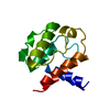
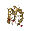
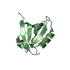
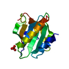
| Title | CALPONIN HOMOLOGY (CH) DOMAIN FROM HUMAN BETA-SPECTRIN AT 1.1 ANGSTROM RESOLUTION | ||||||
 Components Components | SPECTRIN BETA CHAIN | ||||||
 Keywords Keywords | ACTIN-BINDING / FILAMENTOUS ACTIN-BINDING DOMAIN / CYTOSKELETON | ||||||
| Function / homology |  Function and homology information Function and homology informationregulation of SMAD protein signal transduction / membrane assembly / central nervous system formation / spectrin / spectrin-associated cytoskeleton / cuticular plate / plasma membrane organization / Golgi to plasma membrane protein transport / actin filament capping / Nephrin family interactions ...regulation of SMAD protein signal transduction / membrane assembly / central nervous system formation / spectrin / spectrin-associated cytoskeleton / cuticular plate / plasma membrane organization / Golgi to plasma membrane protein transport / actin filament capping / Nephrin family interactions / M band / Sensory processing of sound by outer hair cells of the cochlea / Sensory processing of sound by inner hair cells of the cochlea / Interaction between L1 and Ankyrins / ankyrin binding / RHOV GTPase cycle / cortical actin cytoskeleton / RHOU GTPase cycle / mitotic cytokinesis / axolemma / positive regulation of interleukin-2 production / regulation of protein localization to plasma membrane / COPI-mediated anterograde transport / cell projection / Signaling by FLT3 fusion proteins / NCAM signaling for neurite out-growth / protein localization to plasma membrane / central nervous system development / positive regulation of protein localization to plasma membrane / phospholipid binding / structural constituent of cytoskeleton / actin filament binding / cell junction / RAF/MAP kinase cascade / actin binding / actin cytoskeleton organization / GTPase binding / calmodulin binding / postsynaptic density / cadherin binding / nucleolus / glutamatergic synapse / RNA binding / extracellular exosome / plasma membrane / cytoplasm / cytosol Similarity search - Function | ||||||
| Biological species |  Homo sapiens (human) Homo sapiens (human) | ||||||
| Method |  X-RAY DIFFRACTION / SYNCHROTRON / AB INITIO / Resolution: 1.1 Å X-RAY DIFFRACTION / SYNCHROTRON / AB INITIO / Resolution: 1.1 Å | ||||||
 Authors Authors | Djinovic Carugo, K. / Banuelos, S. / Saraste, M. | ||||||
 Citation Citation | Journal: Structure / Year: 1998 Title: Structural comparisons of calponin homology domains: implications for actin binding. Authors: Banuelos, S. / Saraste, M. / Carugo, K.D. #1: Journal: Nat.Struct.Biol. / Year: 1997Title: Crystal Structure of a Calponin Homology Domain Authors: Carugo, K.D. / Banuelos, S. / Saraste, M. #2: Journal: Nat.Struct.Biol. / Year: 1997Title: The Structure of an Actin-Crosslinking Domain from Human Fimbrin Authors: Goldsmith, S.C. / Pokala, N. / Shen, W. / Fedorov, A.A. / Matsudaira, P. / Almo, S.C. | ||||||
| History |
|
- Structure visualization
| Structure viewer | Molecule: MolmilJmol/JSmol |
|---|
- Downloads & links
-Download
| PDBx/mmCIF format | 1bkr.cif.gz | 67.2 KB | Display | PDBx/mmCIF format |
|---|---|---|---|---|
| PDB format | pdb1bkr.ent.gz | 48.4 KB | Display | PDB format |
| PDBx/mmJSON format | 1bkr.json.gz | Tree view | PDBx/mmJSON format | |
| Others |  Other downloads Other downloads |
-Validation report
| Arichive directory | https://data.pdbj.org/pub/pdb/validation_reports/bk/1bkrftp://data.pdbj.org/pub/pdb/validation_reports/bk/1bkr | HTTPS FTP |
|---|
-Related structure data
| Similar structure data |
|---|


-Links
 PDBj
PDBj


- Assembly
| Deposited unit | 
| ||||||||
|---|---|---|---|---|---|---|---|---|---|
| 1 |
| ||||||||
| Unit cell |
| ||||||||
| Details | SPECTRIN IS A HETEROTETRAMER CONSISTING OF 2 ALPHA AND 2 BETA CHAINS. CALPONIN HOMOLOGY DOMAIN IS AT THE N-TERMINAL PART OF THE BETA CHAIN. |
-Components
| #1: Protein | Mass: 12715.499 Da / Num. of mol.: 1 / Fragment: F-ACTIN BINDING DOMAIN RESIDUES 173 - 281 Source method: isolated from a genetically manipulated source Source: (gene. exp.) Homo sapiens (human) / Cell: NON-ERYTHROCYTE / Cellular location: CYTOSKELETAL PROTEIN / Plasmid: PBAT4 / Species (production host): Escherichia coli / Production host:  |
|---|---|
| #2: Water | ChemComp-HOH /  Mass: 18.015 Da / Num. of mol.: 208 / Source method: isolated from a natural source / Formula: H2O Mass: 18.015 Da / Num. of mol.: 208 / Source method: isolated from a natural source / Formula: H2O |
-Experimental details
-Experiment
| Experiment | Method: X-RAY DIFFRACTION / Number of used crystals: 1 |
|---|
- Sample preparation
| Crystal | Density Matthews: 2.04 Å3/Da / Density % sol: 39.8 % | ||||||||||||||||||||||||||||||||||||||||||||||||||||||
|---|---|---|---|---|---|---|---|---|---|---|---|---|---|---|---|---|---|---|---|---|---|---|---|---|---|---|---|---|---|---|---|---|---|---|---|---|---|---|---|---|---|---|---|---|---|---|---|---|---|---|---|---|---|---|---|
| Crystal grow | pH: 6.6 Details: PROTEIN WAS CRYSTALLIZED FROM 0.2 M SODIUM ACETATE 0.1 M SODIUM CACODYLATE PH 6.6 PEG8K 30% (W/V) | ||||||||||||||||||||||||||||||||||||||||||||||||||||||
| Crystal grow | *PLUS Temperature: 20 ℃ / Method: vapor diffusion / Details: Djinovic, C., (1997) Nat. Struct. Biol., 4, 175. | ||||||||||||||||||||||||||||||||||||||||||||||||||||||
| Components of the solutions | *PLUS
|
-Data collection
| Diffraction | Mean temperature: 100 K |
|---|---|
| Diffraction source | Source: SYNCHROTRON / Site: EMBL/DESY, HAMBURG  / Beamline: BW7B / Wavelength: 0.8883 / Beamline: BW7B / Wavelength: 0.8883 |
| Detector | Type: MARRESEARCH / Detector: IMAGE PLATE / Date: Sep 1, 1996 |
| Radiation | Monochromatic (M) / Laue (L): M / Scattering type: x-ray |
| Radiation wavelength | Wavelength: 0.8883 Å / Relative weight: 1 |
| Reflection | Resolution: 1.1→55 Å / Num. obs: 42236 / % possible obs: 100 % / Observed criterion σ(I): 0 / Redundancy: 4.7 % / Rmerge(I) obs: 0.044 / Net I/σ(I): 12 |
| Reflection shell | Resolution: 1.1→1.11 Å / Rmerge(I) obs: 0.419 / Mean I/σ(I) obs: 2.1 / % possible all: 99.7 |
| Reflection | *PLUS Num. measured all: 199306 |
- Processing
| Software |
| |||||||||||||||||||||||||||||||||
|---|---|---|---|---|---|---|---|---|---|---|---|---|---|---|---|---|---|---|---|---|---|---|---|---|---|---|---|---|---|---|---|---|---|---|
| Refinement | Method to determine structure: AB INITIO / Resolution: 1.1→10 Å / Num. parameters: 9858 / Num. restraintsaints: 11588 / Cross valid method: THROUGHOUT / σ(F): 0 / Stereochemistry target values: ENGH AND HUBER Details: ANISOTROPIC REFINEMENT REDUCED FREE R (NO CUTOFF) FROM 0.225 TO 0.187. ESTIMATED OVERALL COORDINATE ERROR (FROM LUZZATTI PLOT): 0.05 (ASSUMING PERFECT DATA).
| |||||||||||||||||||||||||||||||||
| Solvent computation | Solvent model: MOEWS & KRETSINGER, J.MOL.BIOL.91(1973)201-2 | |||||||||||||||||||||||||||||||||
| Refine analyze | Num. disordered residues: 0 / Occupancy sum hydrogen: 0 / Occupancy sum non hydrogen: 1081.5 | |||||||||||||||||||||||||||||||||
| Refinement step | Cycle: LAST / Resolution: 1.1→10 Å
| |||||||||||||||||||||||||||||||||
| Refine LS restraints |
| |||||||||||||||||||||||||||||||||
| Software | *PLUS Name: SHELXL-97 / Classification: refinement | |||||||||||||||||||||||||||||||||
| Refinement | *PLUS Rfactor Rwork: 0.141 | |||||||||||||||||||||||||||||||||
| Solvent computation | *PLUS | |||||||||||||||||||||||||||||||||
| Displacement parameters | *PLUS |
