 Movie
Movie Controller


+ Open data

- Basic information
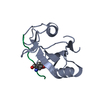
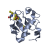
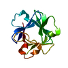
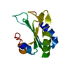
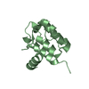
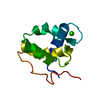
| Entry | Database: PDB / ID: 1aa2 | ||||||
|---|---|---|---|---|---|---|---|
| Title | CALPONIN HOMOLOGY (CH) DOMAIN FROM HUMAN BETA-SPECTRIN | ||||||
 Components Components | BETA-SPECTRIN | ||||||
 Keywords Keywords | CYTOSKELETON / SPECTRIN / F-ACTIN CROSS-LINKING | ||||||
| Function / homology |  Function and homology information Function and homology informationregulation of SMAD protein signal transduction / membrane assembly / central nervous system formation / spectrin / spectrin-associated cytoskeleton / cuticular plate / plasma membrane organization / Golgi to plasma membrane protein transport / actin filament capping / Nephrin family interactions ...regulation of SMAD protein signal transduction / membrane assembly / central nervous system formation / spectrin / spectrin-associated cytoskeleton / cuticular plate / plasma membrane organization / Golgi to plasma membrane protein transport / actin filament capping / Nephrin family interactions / M band / Sensory processing of sound by outer hair cells of the cochlea / Sensory processing of sound by inner hair cells of the cochlea / Interaction between L1 and Ankyrins / ankyrin binding / RHOV GTPase cycle / cortical actin cytoskeleton / RHOU GTPase cycle / mitotic cytokinesis / axolemma / positive regulation of interleukin-2 production / regulation of protein localization to plasma membrane / COPI-mediated anterograde transport / cell projection / Signaling by FLT3 fusion proteins / NCAM signaling for neurite out-growth / protein localization to plasma membrane / positive regulation of protein localization to plasma membrane / central nervous system development / phospholipid binding / structural constituent of cytoskeleton / actin filament binding / cell junction / RAF/MAP kinase cascade / actin binding / actin cytoskeleton organization / GTPase binding / calmodulin binding / postsynaptic density / cadherin binding / nucleolus / glutamatergic synapse / RNA binding / extracellular exosome / plasma membrane / cytoplasm / cytosol Similarity search - Function | ||||||
| Biological species |  Homo sapiens (human) Homo sapiens (human) | ||||||
| Method |  X-RAY DIFFRACTION / MIR / Resolution: 2 Å X-RAY DIFFRACTION / MIR / Resolution: 2 Å | ||||||
 Authors Authors | Djinovic Carugo, K. / Banuelos, S. / Saraste, M. | ||||||
 Citation Citation | Journal: Nat.Struct.Biol. / Year: 1997 Title: Crystal structure of a calponin homology domain. Authors: Djinovic Carugo, K. / Banuelos, S. / Saraste, M. #1: Journal: J.Biol.Chem. / Year: 1996Title: Identification of a Phosphatidylinositol 4,5-Bisphosphate-Binding Site in Chicken Skeletal Muscle Alpha-Actinin Authors: Fukami, K. / Sawada, N. / Endo, T. / Takenawa, T. #2: Journal: FEBS Lett. / Year: 1995Title: Does Vav Bind to F-Actin Through a Ch Domain? Authors: Castresana, J. / Saraste, M. #3: Journal: Protein Profile / Year: 1994Title: Actin-Binding Proteins 1: Spectrin Superfamily Authors: Hartwig, J.H. | ||||||
| History |
|
- Structure visualization
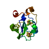
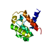
| Structure viewer | Molecule: MolmilJmol/JSmol |
|---|
- Downloads & links
-Download
| PDBx/mmCIF format | 1aa2.cif.gz | 38.5 KB | Display | PDBx/mmCIF format |
|---|---|---|---|---|
| PDB format | pdb1aa2.ent.gz | 24.9 KB | Display | PDB format |
| PDBx/mmJSON format | 1aa2.json.gz | Tree view | PDBx/mmJSON format | |
| Others |  Other downloads Other downloads |
-Validation report
| Arichive directory | https://data.pdbj.org/pub/pdb/validation_reports/aa/1aa2ftp://data.pdbj.org/pub/pdb/validation_reports/aa/1aa2 | HTTPS FTP |
|---|
-Related structure data
| Similar structure data |
|---|


-Links
 PDBj
PDBj


- Assembly
| Deposited unit | 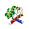
| ||||||||
|---|---|---|---|---|---|---|---|---|---|
| 1 |
| ||||||||
| Unit cell |
|
-Components
| #1: Protein | Mass: 12586.320 Da / Num. of mol.: 1 / Fragment: F-ACTIN BINDING DOMAIN RESIDUES 173 - 281 Source method: isolated from a genetically manipulated source Source: (gene. exp.) Homo sapiens (human) / Organelle: NON-ERYTHROCYTE / Plasmid: PBAT4 / Species (production host): Escherichia coli / Production host:  |
|---|---|
| #2: Water | ChemComp-HOH /  Mass: 18.015 Da / Num. of mol.: 130 / Source method: isolated from a natural source / Formula: H2O Mass: 18.015 Da / Num. of mol.: 130 / Source method: isolated from a natural source / Formula: H2O |
-Experimental details
-Experiment
| Experiment | Method: X-RAY DIFFRACTION / Number of used crystals: 1 |
|---|
- Sample preparation
| Crystal | Density Matthews: 2.04 Å3/Da / Density % sol: 39.8 % | ||||||||||||||||||||||||||||||||||||||||||||||||||||||
|---|---|---|---|---|---|---|---|---|---|---|---|---|---|---|---|---|---|---|---|---|---|---|---|---|---|---|---|---|---|---|---|---|---|---|---|---|---|---|---|---|---|---|---|---|---|---|---|---|---|---|---|---|---|---|---|
| Crystal grow | pH: 6.6 Details: PROTEIN WAS CRYSTALLIZED FROM 30% PEG 8000, 100 MM SODIUM CACODYLATE, PH 6.6 | ||||||||||||||||||||||||||||||||||||||||||||||||||||||
| Crystal grow | *PLUS Temperature: 20 ℃ / Method: vapor diffusion | ||||||||||||||||||||||||||||||||||||||||||||||||||||||
| Components of the solutions | *PLUS
|
-Data collection
| Diffraction | Mean temperature: 100 K |
|---|---|
| Diffraction source | Source: ROTATING ANODE / Type: ENRAF-NONIUS FR571 / Wavelength: 1.5418 |
| Detector | Type: MARRESEARCH / Detector: IMAGE PLATE / Date: Feb 1, 1996 / Details: NO OPTICS |
| Radiation | Monochromator: GRAPHITE(002) / Monochromatic (M) / Laue (L): M / Scattering type: x-ray |
| Radiation wavelength | Wavelength: 1.5418 Å / Relative weight: 1 |
| Reflection | Resolution: 2→14 Å / Num. obs: 6859 / % possible obs: 99.1 % / Observed criterion σ(I): 0 / Redundancy: 5.5 % / Biso Wilson estimate: 21.5 Å2 / Rmerge(I) obs: 0.074 / Rsym value: 0.08 / Net I/σ(I): 12 |
| Reflection shell | Resolution: 2.01→2.04 Å / Redundancy: 3 % / Rmerge(I) obs: 0.265 / Mean I/σ(I) obs: 3 / Rsym value: 0.311 / % possible all: 97.2 |
| Reflection | *PLUS Num. measured all: 27400 |
- Processing
| Software |
| ||||||||||||||||||||||||||||||||||||||||||||||||||
|---|---|---|---|---|---|---|---|---|---|---|---|---|---|---|---|---|---|---|---|---|---|---|---|---|---|---|---|---|---|---|---|---|---|---|---|---|---|---|---|---|---|---|---|---|---|---|---|---|---|---|---|
| Refinement | Method to determine structure: MIR / Resolution: 2→14 Å / Isotropic thermal model: TNT BCORREL / Cross valid method: A POSTERIORI / σ(F): 0 / Stereochemistry target values: TNT PROTGEO / Details: THE REPORTED RMS VALUES ARE UNWEIGHTED
| ||||||||||||||||||||||||||||||||||||||||||||||||||
| Solvent computation | Solvent model: MOEWS & KRETSINGER (1975) J. MOL. BIOL. 91, 201-228. Bsol: 83.74 Å2 / ksol: 0.754 e/Å3 | ||||||||||||||||||||||||||||||||||||||||||||||||||
| Refinement step | Cycle: LAST / Resolution: 2→14 Å
| ||||||||||||||||||||||||||||||||||||||||||||||||||
| Refine LS restraints |
| ||||||||||||||||||||||||||||||||||||||||||||||||||
| Software | *PLUS Name: TNT / Version: 5EA / Classification: refinement | ||||||||||||||||||||||||||||||||||||||||||||||||||
| Refinement | *PLUS Rfactor all: 0.16 / Rfactor Rfree: 0.213 / Rfactor Rwork: 0.16 | ||||||||||||||||||||||||||||||||||||||||||||||||||
| Solvent computation | *PLUS | ||||||||||||||||||||||||||||||||||||||||||||||||||
| Displacement parameters | *PLUS | ||||||||||||||||||||||||||||||||||||||||||||||||||
| Refine LS restraints | *PLUS
|
