[English] 日本語
Yorodumi
- PDB-2v2k: THE CRYSTAL STRUCTURE OF FDXA, A 7FE FERREDOXIN FROM MYCOBACTERIU... -

+
Open data


ID or keywords:

Loading...

-
Basic information

Entry
Database: PDB / ID: 2v2k
Title
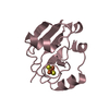
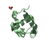
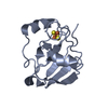
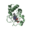
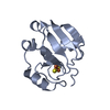
THE CRYSTAL STRUCTURE OF FDXA, A 7FE FERREDOXIN FROM MYCOBACTERIUM SMEGMATIS
ComponentsFERREDOXIN
KeywordsELECTRON TRANSPORT / IRON / TRANSPORT / IRON-SULFUR / MYCOBACTERIUM TUBERCULOSIS / FE-S CLUSTER / METAL-BINDING / 7FE FERREDOXIN / ELECTRON TRANSFER / MYCOBACTERIUM SMEGMATIS / 4FE-4S CLUSTER INSTABILITY
Function / homology
Function and homology information


3 iron, 4 sulfur cluster binding / 4 iron, 4 sulfur cluster binding / electron transfer activity / metal ion binding
Similarity search - Function
7Fe ferredoxin / 4Fe-4S binding domain / Alpha-Beta Plaits - #20 / 4Fe-4S ferredoxin, iron-sulphur binding, conserved site / 4Fe-4S ferredoxin-type iron-sulfur binding region signature. / 4Fe-4S ferredoxin-type iron-sulfur binding domain profile. / 4Fe-4S ferredoxin-type, iron-sulphur binding domain / Alpha-Beta Plaits / 2-Layer Sandwich / Alpha Beta
Similarity search - Domain/homology
ACETATE ION / FE3-S4 CLUSTER / Ferredoxin
Similarity search - Component
Biological speciesMYCOBACTERIUM SMEGMATIS (bacteria)
MethodX-RAY DIFFRACTION / SYNCHROTRON / MOLECULAR REPLACEMENT / Resolution: 1.6 Å
AuthorsRicagno, S. / de Rosa, M. / Aliverti, A. / Zanetti, G. / Bolognesi, M.
CitationJournal: Biochem.Biophys.Res.Commun. / Year: 2007
Title: The Crystal Structure of Fdxa, a 7Fe Ferredoxin from Mycobacterium Smegmatis.
Authors: Ricagno, S. / De Rosa, M. / Aliverti, A. / Zanetti, G. / Bolognesi, M.
History
DepositionJun 6, 2007Deposition site: PDBE / Processing site: PDBE
Revision 1.0Jul 3, 2007Provider: repository / Type: Initial release
Revision 1.1Jul 13, 2011Group: Advisory / Version format compliance
Revision 1.2Dec 13, 2023Group: Data collection / Database references ...Data collection / Database references / Derived calculations / Other / Refinement description
Category: chem_comp_atom / chem_comp_bond ...chem_comp_atom / chem_comp_bond / database_2 / pdbx_database_status / pdbx_initial_refinement_model / pdbx_struct_conn_angle / pdbx_struct_special_symmetry / struct_conn / struct_site
Item: _database_2.pdbx_DOI / _database_2.pdbx_database_accession ..._database_2.pdbx_DOI / _database_2.pdbx_database_accession / _pdbx_database_status.status_code_sf / _pdbx_struct_conn_angle.ptnr1_auth_seq_id / _pdbx_struct_conn_angle.ptnr1_label_asym_id / _pdbx_struct_conn_angle.ptnr1_label_atom_id / _pdbx_struct_conn_angle.ptnr1_label_seq_id / _pdbx_struct_conn_angle.ptnr2_auth_seq_id / _pdbx_struct_conn_angle.ptnr2_label_asym_id / _pdbx_struct_conn_angle.ptnr2_label_atom_id / _pdbx_struct_conn_angle.ptnr3_auth_seq_id / _pdbx_struct_conn_angle.ptnr3_label_asym_id / _pdbx_struct_conn_angle.ptnr3_label_atom_id / _pdbx_struct_conn_angle.value / _struct_conn.conn_type_id / _struct_conn.id / _struct_conn.pdbx_dist_value / _struct_conn.pdbx_leaving_atom_flag / _struct_conn.ptnr1_auth_asym_id / _struct_conn.ptnr1_auth_comp_id / _struct_conn.ptnr1_auth_seq_id / _struct_conn.ptnr1_label_asym_id / _struct_conn.ptnr1_label_atom_id / _struct_conn.ptnr1_label_comp_id / _struct_conn.ptnr1_label_seq_id / _struct_conn.ptnr2_auth_asym_id / _struct_conn.ptnr2_auth_comp_id / _struct_conn.ptnr2_auth_seq_id / _struct_conn.ptnr2_label_asym_id / _struct_conn.ptnr2_label_atom_id / _struct_conn.ptnr2_label_comp_id / _struct_conn.ptnr2_label_seq_id / _struct_site.pdbx_auth_asym_id / _struct_site.pdbx_auth_comp_id / _struct_site.pdbx_auth_seq_id

-
Structure visualization

Structure viewerMolecule:
MolmilJmol/JSmol

Downloads & links

-
Assembly

Deposited unit
A: FERREDOXIN
B: FERREDOXIN
hetero molecules


Theoretical massNumber of molelcules
Total (without water)24,1077
Polymers22,8652
Non-polymers1,2425
Water2,126118
1
A: FERREDOXIN
hetero molecules


Theoretical massNumber of molelcules
Total (without water)12,0243
Polymers11,4331
Non-polymers5922
Water181
TypeNameSymmetry operationNumber
identity operation1_555x,y,z1
MethodPQS
2
B: FERREDOXIN
hetero molecules


Theoretical massNumber of molelcules
Total (without water)12,0834
Polymers11,4331
Non-polymers6513
Water181
TypeNameSymmetry operationNumber
identity operation1_555x,y,z1
MethodPQS
Unit cell
Length a, b, c (Å)54.581, 54.526, 135.382
Angle α, β, γ (deg.)90.00, 90.00, 90.00
Int Tables number20
Space group name H-MC2221
Components on special symmetry positions
IDModelComponents
11A-2005-

HOH

-
Components

#1: Protein FERREDOXIN / / 7-IRON FERREDOXIN


Mass: 11432.598 Da / Num. of mol.: 2 / Fragment: RESIDUES 1-105 / Source method: isolated from a natural source / Source: (natural) MYCOBACTERIUM SMEGMATIS (bacteria) / Strain: MC2 / References: UniProt: P00215
#2: Chemical
ChemComp-F3S / FE3-S4 CLUSTER / Iron–sulfur cluster


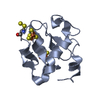
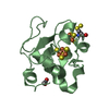
Mass: 295.795 Da / Num. of mol.: 4 / Source method: obtained synthetically / Formula: Fe3S4
#3: Chemical ChemComp-ACT / ACETATE ION / Acetate


Mass: 59.044 Da / Num. of mol.: 1 / Source method: obtained synthetically / Formula: C2H3O2
#4: Water ChemComp-HOH / water / Water


Mass: 18.015 Da / Num. of mol.: 118 / Source method: isolated from a natural source / Formula: H2O
Compound detailsFERREDOXINS ARE IRON-SULFUR PROTEINS THAT TRANSFER ELECTRONS IN A WIDE VARIETY OF METABOLIC REACTIONS.

-
Experimental details

-
Experiment

ExperimentMethod: X-RAY DIFFRACTION / Number of used crystals: 1

-
Sample preparation

CrystalDensity Matthews: 2.28 Å3/Da / Density % sol: 46 %
Crystal growpH: 5.5 / Details: AMMONIUM SULPHATE, MPD, SODIUM ACETATE, pH 5.50

-
Data collection

DiffractionMean temperature: 110 K
Diffraction sourceSource: SYNCHROTRON / Site: ESRF / Beamline: ID14-1 / Wavelength: 0.931
DetectorType: ADSC CCD / Detector: CCD / Date: Apr 26, 2007
RadiationMonochromator: DIAMONDS CRYSTALS / Protocol: SINGLE WAVELENGTH / Monochromatic (M) / Laue (L): M / Scattering type: x-ray
Radiation wavelengthWavelength: 0.931 Å / Relative weight: 1
ReflectionResolution: 1.6→20 Å / Num. obs: 26692 / % possible obs: 93.9 % / Observed criterion σ(I): 2 / Redundancy: 3.5 % / Rmerge(I) obs: 0.07 / Net I/σ(I): 12.9
Reflection shellResolution: 1.6→1.69 Å / Redundancy: 3.5 % / Rmerge(I) obs: 0.34 / Mean I/σ(I) obs: 2.7 / % possible all: 99.9

-
Processing

Software
NameVersionClassification
REFMAC5.2.0019refinement
MOSFLMdata reduction
MOSFLMdata scaling
MOLREPphasing
RefinementMethod to determine structure: MOLECULAR REPLACEMENT
Starting model: PDB ENTRY 1H98
Resolution: 1.6→67.73 Å / Cor.coef. Fo:Fc: 0.966 / Cor.coef. Fo:Fc free: 0.946 / SU B: 4.047 / SU ML: 0.067 / TLS residual ADP flag: LIKELY RESIDUAL / Cross valid method: THROUGHOUT / ESU R: 0.083 / ESU R Free: 0.087 / Stereochemistry target values: MAXIMUM LIKELIHOOD
Details: HYDROGENS HAVE BEEN ADDED IN THE RIDING POSITIONS. THIS ENTRY CONTAINS SOME ATOMS REFINED WITH ZERO OCCUPANCY FOR WHICH B-FACTORS HAVE BEEN REFINED.
RfactorNum. reflection% reflectionSelection details
Rfree0.202 1344 5 %RANDOM
Rwork0.165 ---
obs0.167 25306 98.4 %-
Solvent computationIon probe radii: 0.8 Å / Shrinkage radii: 0.8 Å / VDW probe radii: 1.4 Å / Solvent model: MASK
Displacement parametersBiso mean: 18.54 Å2
Baniso -1Baniso -2Baniso -3
1--0.67 Å20 Å20 Å2
2--2.49 Å20 Å2
3----1.82 Å2
Refinement stepCycle: LAST / Resolution: 1.6→67.73 Å
ProteinNucleic acidLigandSolventTotal
Num. atoms1569 0 32 118 1719
Refine LS restraints
Refine-IDTypeDev idealDev ideal targetNumber
X-RAY DIFFRACTIONr_bond_refined_d0.0160.0221676
X-RAY DIFFRACTIONr_bond_other_d0.0180.021062
X-RAY DIFFRACTIONr_angle_refined_deg1.5282.0052293
X-RAY DIFFRACTIONr_angle_other_deg1.22332633
X-RAY DIFFRACTIONr_dihedral_angle_1_deg6.0645212
X-RAY DIFFRACTIONr_dihedral_angle_2_deg38.87626.88377
X-RAY DIFFRACTIONr_dihedral_angle_3_deg13.51215239
X-RAY DIFFRACTIONr_dihedral_angle_4_deg7.664152
X-RAY DIFFRACTIONr_chiral_restr0.7070.2250
X-RAY DIFFRACTIONr_gen_planes_refined0.0060.021867
X-RAY DIFFRACTIONr_gen_planes_other0.0010.02277
X-RAY DIFFRACTIONr_nbd_refined0.2980.2393
X-RAY DIFFRACTIONr_nbd_other0.1840.21086
X-RAY DIFFRACTIONr_nbtor_refined0.1670.2814
X-RAY DIFFRACTIONr_nbtor_other0.0860.2650
X-RAY DIFFRACTIONr_xyhbond_nbd_refined0.1780.276
X-RAY DIFFRACTIONr_xyhbond_nbd_other
X-RAY DIFFRACTIONr_metal_ion_refined
X-RAY DIFFRACTIONr_metal_ion_other
X-RAY DIFFRACTIONr_symmetry_vdw_refined0.1240.23
X-RAY DIFFRACTIONr_symmetry_vdw_other0.1920.219
X-RAY DIFFRACTIONr_symmetry_hbond_refined0.1740.27
X-RAY DIFFRACTIONr_symmetry_hbond_other
X-RAY DIFFRACTIONr_symmetry_metal_ion_refined
X-RAY DIFFRACTIONr_symmetry_metal_ion_other
X-RAY DIFFRACTIONr_mcbond_it1.131.51356
X-RAY DIFFRACTIONr_mcbond_other
X-RAY DIFFRACTIONr_mcangle_it1.12821716
X-RAY DIFFRACTIONr_mcangle_other
X-RAY DIFFRACTIONr_scbond_it2.2233718
X-RAY DIFFRACTIONr_scbond_other
X-RAY DIFFRACTIONr_scangle_it3.1644.5552
X-RAY DIFFRACTIONr_scangle_other
X-RAY DIFFRACTIONr_long_range_B_refined
X-RAY DIFFRACTIONr_long_range_B_other
X-RAY DIFFRACTIONr_rigid_bond_restr
X-RAY DIFFRACTIONr_sphericity_free
X-RAY DIFFRACTIONr_sphericity_bonded
LS refinement shellResolution: 1.6→1.64 Å / Total num. of bins used: 20 /
RfactorNum. reflection
Rfree0.289 105
Rwork0.261 1839
Refinement TLS params.

Method: refined / Refine-ID: X-RAY DIFFRACTION

IDL112)L122)L132)L222)L232)L332)S11 (Å °)S12 (Å °)S13 (Å °)S21 (Å °)S22 (Å °)S23 (Å °)S31 (Å °)S32 (Å °)S33 (Å °)T112)T122)T132)T222)T232)T332)Origin x (Å)Origin y (Å)Origin z (Å)
12.93540.9577-0.70510.8802-0.50774.5215-0.0195-0.1101-0.02550.0621-0.0552-0.06480.01880.02490.0746-0.06030.0043-0.0058-0.05210.0153-0.072720.056211.908722.1878
24.23140.7602-1.07071.1494-0.7233.7401-0.193-0.1445-0.4257-0.05250.0423-0.10320.4335-0.26170.1507-0.0103-0.04410.0265-0.06480.016-0.051315.55085.044821.6034
32.294-0.50511.06582.1459-2.07077.40290.1685-0.3750.0707-0.029-0.1229-0.08750.2385-0.4538-0.0456-0.0707-0.0310.02880.10450.0279-0.088810.82648.695932.5961
40.089-0.605-0.32684.14682.19261.2225-0.15020.07150.13540.0271-0.1534-0.18240.1432-0.22360.30360.0116-0.0089-0.00280.02490.01540.032321.48311.597514.631
516.5168-5.2046-9.8012.03424.837913.5818-0.1277-0.33310.1226-0.06230.0754-0.0102-0.12330.22330.05220.0192-0.0141-0.01490.03150.0054-0.001222.264714.301526.3929
61.9592-0.1871-0.37331.019-0.44772.4068-0.02330.0315-0.0755-0.04810.00970.00160.0789-0.07790.0136-0.0446-0.00380.0019-0.0701-0.0091-0.036219.565813.6859-7.5864
73.05210.3036-1.23270.931-0.3852.1650.08020.03270.25080.06590.0250.0035-0.2328-0.1169-0.1052-0.03460.02430.0102-0.0670.0068-0.026518.824622.4069-6.9487
80.66461.281-0.86362.5734-2.33545.4371-0.00210.1759-0.0292-0.12090.04330.1093-0.1045-0.224-0.0412-0.06710.0215-0.02020.04930.0246-0.036211.072519.9256-15.8261
98.6508-3.43840.2437.87260.58060.0773-0.10170.1135-0.12830.1129-0.0906-0.0185-0.07350.04290.19240.0174-0.00490.0130.00080.01650.034421.401313.2401-0.1249
1028.9756-7.798619.62072.356-3.823621.54590.10740.2632-0.39620.2189-0.0099-0.05770.0211-0.0345-0.09740.0094-0.00850.0065-0.0614-0.02310.023420.329510.7948-11.8632
Refinement TLS group
IDRefine-IDRefine TLS-IDAuth asym-IDAuth seq-ID
1X-RAY DIFFRACTION1A1 - 55
2X-RAY DIFFRACTION2A56 - 86
3X-RAY DIFFRACTION3A87 - 104
4X-RAY DIFFRACTION4A107
5X-RAY DIFFRACTION5A108
6X-RAY DIFFRACTION6B1 - 55
7X-RAY DIFFRACTION7B56 - 85
8X-RAY DIFFRACTION8B86 - 103
9X-RAY DIFFRACTION9B107
10X-RAY DIFFRACTION10B108

+
About Yorodumi

-
News

-
Feb 9, 2022. New format data for meta-information of EMDB entries

New format data for meta-information of EMDB entries

  • Version 3 of the EMDB header file is now the official format.
  • The previous official version 1.9 will be removed from the archive.

Related info.:EMDB header

External links:wwPDB to switch to version 3 of the EMDB data model

-
Aug 12, 2020. Covid-19 info

Covid-19 info

URL: https://pdbj.org/emnavi/covid19.php

New page: Covid-19 featured information page in EM Navigator.

Related info.:Covid-19 info / Mar 5, 2020. Novel coronavirus structure data

+
Mar 5, 2020. Novel coronavirus structure data

Novel coronavirus structure data

Related info.:Yorodumi Speices / Aug 12, 2020. Covid-19 info

External links:COVID-19 featured content - PDBj / Molecule of the Month (242):Coronavirus Proteases

+
Jan 31, 2019. EMDB accession codes are about to change! (news from PDBe EMDB page)

EMDB accession codes are about to change! (news from PDBe EMDB page)

  • The allocation of 4 digits for EMDB accession codes will soon come to an end. Whilst these codes will remain in use, new EMDB accession codes will include an additional digit and will expand incrementally as the available range of codes is exhausted. The current 4-digit format prefixed with “EMD-” (i.e. EMD-XXXX) will advance to a 5-digit format (i.e. EMD-XXXXX), and so on. It is currently estimated that the 4-digit codes will be depleted around Spring 2019, at which point the 5-digit format will come into force.
  • The EM Navigator/Yorodumi systems omit the EMD- prefix.

Related info.:Q: What is EMD? / ID/Accession-code notation in Yorodumi/EM Navigator

External links:EMDB Accession Codes are Changing Soon! / Contact to PDBj

+
Jul 12, 2017. Major update of PDB

Major update of PDB

  • wwPDB released updated PDB data conforming to the new PDBx/mmCIF dictionary.
  • This is a major update changing the version number from 4 to 5, and with Remediation, in which all the entries are updated.
  • In this update, many items about electron microscopy experimental information are reorganized (e.g. em_software).
  • Now, EM Navigator and Yorodumi are based on the updated data.

External links:wwPDB Remediation / Enriched Model Files Conforming to OneDep Data Standards Now Available in the PDB FTP Archive

-
Yorodumi

Thousand views of thousand structures

  • Yorodumi is a browser for structure data from EMDB, PDB, SASBDB, etc.
  • This page is also the successor to EM Navigator detail page, and also detail information page/front-end page for Omokage search.
  • The word "yorodu" (or yorozu) is an old Japanese word meaning "ten thousand". "mi" (miru) is to see.

Related info.:EMDB / PDB / SASBDB / Comparison of 3 databanks / Yorodumi Search / Aug 31, 2016. New EM Navigator & Yorodumi / Yorodumi Papers / Jmol/JSmol / Function and homology information / Changes in new EM Navigator and Yorodumi

Read more