 Movie
Movie Controller


+ Open data

- Basic information
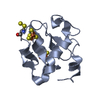
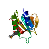
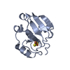
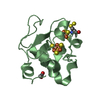
| Entry | Database: PDB / ID: 1a75 | ||||||
|---|---|---|---|---|---|---|---|
| Title | WHITING PARVALBUMIN | ||||||
 Components Components | (PARVALBUMIN) x 2 | ||||||
 Keywords Keywords | CALCIUM BINDING PROTEIN / MUSCLE PROTEIN | ||||||
| Function / homology |  Function and homology information Function and homology information | ||||||
| Biological species |  Merlangius merlangus (whiting) Merlangius merlangus (whiting) | ||||||
| Method |  X-RAY DIFFRACTION / SYNCHROTRON / MOLECULAR REPLACEMENT / Resolution: 1.9 Å X-RAY DIFFRACTION / SYNCHROTRON / MOLECULAR REPLACEMENT / Resolution: 1.9 Å | ||||||
 Authors Authors | Declercq, J.P. / Baneres, J.L. / Rambaud, J. / Parello, J. | ||||||
 Citation Citation | Journal: To be Published Title: Tertiary Structure of a Trp-Containing Parvalbumin from Whiting (Merlangius Merlangus). Description of the Hydrophobic Core Authors: Declercq, J.P. / Baneres, J.L. / Rambaud, J. / Parello, J. #1: Journal: Acta Crystallogr.,Sect.D / Year: 1996Title: X-Ray Structure of a New Crystal Form of Pike 4.10 Beta Parvalbumin Authors: Declercq, J.P. / Tinant, B. / Parello, J. #2: Journal: J.Mol.Biol. / Year: 1992Title: Crystal Structure of the Unique Parvalbumin Component from Muscle of the Leopard Shark (Triakis Semifasciata). The First X-Ray Study of an Alpha-Parvalbumin Authors: Roquet, F. / Declercq, J.P. / Tinant, B. / Rambaud, J. / Parello, J. #3: Journal: J.Mol.Biol. / Year: 1991Title: Ionic Interactions with Parvalbumins. Crystal Structure Determination of Pike 4.10 Parvalbumin in Four Different Ionic Environments Authors: Declercq, J.P. / Tinant, B. / Parello, J. / Rambaud, J. #4: Journal: J.Mol.Biol. / Year: 1988Title: Crystal Structure Determination and Refinement of Pike 4.10 Parvalbumin (Minor Component from Esox Lucius) Authors: Declercq, J.P. / Tinant, B. / Parello, J. / Etienne, G. / Huber, R. | ||||||
| History |
|
- Structure visualization
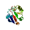
| Structure viewer | Molecule: MolmilJmol/JSmol |
|---|
- Downloads & links
-Download
| PDBx/mmCIF format | 1a75.cif.gz | 59.1 KB | Display | PDBx/mmCIF format |
|---|---|---|---|---|
| PDB format | pdb1a75.ent.gz | 41.8 KB | Display | PDB format |
| PDBx/mmJSON format | 1a75.json.gz | Tree view | PDBx/mmJSON format | |
| Others |  Other downloads Other downloads |
-Validation report
| Arichive directory | https://data.pdbj.org/pub/pdb/validation_reports/a7/1a75ftp://data.pdbj.org/pub/pdb/validation_reports/a7/1a75 | HTTPS FTP |
|---|
-Related structure data
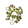
| Related structure data | 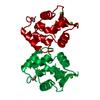 5cpvS S: Starting model for refinement |
|---|---|
| Similar structure data |
-Links
 PDBj
PDBj- Assembly
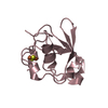
| Deposited unit | 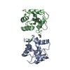
| ||||||||
|---|---|---|---|---|---|---|---|---|---|
| 1 | 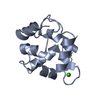
| ||||||||
| 2 | 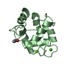
| ||||||||
| Unit cell |
| ||||||||
| Noncrystallographic symmetry (NCS) | NCS oper: (Code: given Matrix: (0.9998, -0.0184, 0.0012), Vector: |
-Components
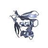
| #1: Protein | Mass: 11335.788 Da / Num. of mol.: 1 / Source method: isolated from a natural source / Source: (natural) Merlangius merlangus (whiting) / Tissue: MUSCLE / References: UniProt: P02621 | ||||
|---|---|---|---|---|---|
| #2: Protein | Mass: 11361.825 Da / Num. of mol.: 1 / Source method: isolated from a natural source / Source: (natural) Merlangius merlangus (whiting) / Tissue: MUSCLE / References: UniProt: P02621 | ||||
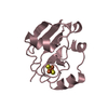
| #3: Chemical | ChemComp-CA /   Mass: 40.078 Da / Num. of mol.: 4 / Source method: obtained synthetically / Formula: Ca Mass: 40.078 Da / Num. of mol.: 4 / Source method: obtained synthetically / Formula: Ca#4: Water | ChemComp-HOH / |  Mass: 18.015 Da / Num. of mol.: 235 / Source method: isolated from a natural source / Formula: H2O Mass: 18.015 Da / Num. of mol.: 235 / Source method: isolated from a natural source / Formula: H2OHas protein modification | Y | |
-Experimental details
-Experiment
| Experiment | Method: X-RAY DIFFRACTION / Number of used crystals: 1 |
|---|
- Sample preparation
| Crystal | Density Matthews: 2.08 Å3/Da / Density % sol: 41 % |
|---|---|
| Crystal grow | Method: vapor diffusion, sitting drop / pH: 6 Details: SITTING DROP. RESERVOIR: 1 ML 2.1M NAH2PO4/NA2HPO4 (PH 6.0) 0.7M (NH4)2SO4, 0.02%(W/V) NAN3 DROP: 10 MICROL. PROTEIN SOLUTION (15MG/ML) +10 MICROL. RESERVOIR SOLUTION., vapor diffusion - sitting drop |
-Data collection
| Diffraction | Mean temperature: 100 K |
|---|---|
| Diffraction source | Source: SYNCHROTRON / Site: EMBL/DESY, HAMBURG  / Beamline: BW7A / Wavelength: 0.9 / Beamline: BW7A / Wavelength: 0.9 |
| Detector | Type: MARRESEARCH / Detector: IMAGE PLATE / Date: Sep 1, 1996 / Details: FOCUSING MIRROR |
| Radiation | Monochromator: DOUBLE CRYSTAL SI(111) / Monochromatic (M) / Laue (L): M / Scattering type: x-ray |
| Radiation wavelength | Wavelength: 0.9 Å / Relative weight: 1 |
| Reflection | Resolution: 1.9→12 Å / Num. obs: 12412 / % possible obs: 82.2 % / Redundancy: 1.54 % / Rsym value: 0.057 / Net I/σ(I): 8.6 |
| Reflection shell | Resolution: 1.9→1.93 Å / Redundancy: 1.48 % / Mean I/σ(I) obs: 5 / Rsym value: 0.12 / % possible all: 73.2 |
- Processing
| Software |
| |||||||||||||||||||||||||||||||||
|---|---|---|---|---|---|---|---|---|---|---|---|---|---|---|---|---|---|---|---|---|---|---|---|---|---|---|---|---|---|---|---|---|---|---|
| Refinement | Method to determine structure: MOLECULAR REPLACEMENT Starting model: PDB ENTRY 5CPV Resolution: 1.9→12 Å / Num. parameters: 7243 / Num. restraintsaints: 7757 / σ(F): 0 / Stereochemistry target values: ENGH AND HUBER
| |||||||||||||||||||||||||||||||||
| Solvent computation | Solvent model: MOEWS & KRETSINGER | |||||||||||||||||||||||||||||||||
| Refine analyze | Num. disordered residues: 0 / Occupancy sum hydrogen: 0 / Occupancy sum non hydrogen: 1810 | |||||||||||||||||||||||||||||||||
| Refinement step | Cycle: LAST / Resolution: 1.9→12 Å
| |||||||||||||||||||||||||||||||||
| Refine LS restraints |
|
