 Movie
Movie Controller

[English] 日本語
 Yorodumi
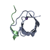
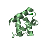
- PDB-1h98: New Insights into Thermostability of Bacterial Ferredoxins: High ... -
+ Open data

- Basic information
| Entry | Database: PDB / ID: 1h98 | ||||||
|---|---|---|---|---|---|---|---|
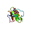
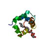
| Title | New Insights into Thermostability of Bacterial Ferredoxins: High Resolution Crystal Structure of the Seven-Iron Ferredoxin from Thermus thermophilus | ||||||
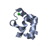
 Components Components | FERREDOXIN | ||||||
 Keywords Keywords | ELECTRON TRANSPORT / THERMOPHILIC / IRON-SULFUR / AZOTOBACTER / HYDROGEN BONDS / STABILITY / HIGH RESOLUTION | ||||||
| Function / homology |  Function and homology information Function and homology information3 iron, 4 sulfur cluster binding / 4 iron, 4 sulfur cluster binding / electron transfer activity / metal ion binding Similarity search - Function | ||||||
| Biological species |   THERMUS AQUATICUS (bacteria) THERMUS AQUATICUS (bacteria) | ||||||
| Method |  X-RAY DIFFRACTION / MOLECULAR REPLACEMENT / Resolution: 1.64 Å X-RAY DIFFRACTION / MOLECULAR REPLACEMENT / Resolution: 1.64 Å | ||||||
 Authors Authors | Macedo-Ribeiro, S. / Martins, B.M. / Pereira, P.J.B. / Buse, G. / Huber, R. / Soulimane, T. | ||||||
 Citation Citation | Journal: J.Biol.Inorg.Chem. / Year: 2001 Title: New Insights Into the Thermostability of Bacterial Ferredoxins: High-Resolution Crystal Structure of the Seven-Iron Ferredoxin from Thermus Thermophilus Authors: Macedo-Ribeiro, S. / Martins, B.M. / Pereira, P.J.B. / Buse, G. / Huber, R. / Soulimane, T. | ||||||
| History |
|
- Structure visualization
| Structure viewer | Molecule: MolmilJmol/JSmol |
|---|
- Downloads & links
-Download
| PDBx/mmCIF format | 1h98.cif.gz | 29.9 KB | Display | PDBx/mmCIF format |
|---|---|---|---|---|
| PDB format | pdb1h98.ent.gz | 18.6 KB | Display | PDB format |
| PDBx/mmJSON format | 1h98.json.gz | Tree view | PDBx/mmJSON format | |
| Others |  Other downloads Other downloads |
-Validation report
| Arichive directory | https://data.pdbj.org/pub/pdb/validation_reports/h9/1h98ftp://data.pdbj.org/pub/pdb/validation_reports/h9/1h98 | HTTPS FTP |
|---|
-Related structure data
| Related structure data | 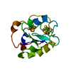 6fd1S S: Starting model for refinement |
|---|---|
| Similar structure data |


-Links
 PDBj
PDBj


- Assembly
| Deposited unit | 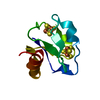
| ||||||||
|---|---|---|---|---|---|---|---|---|---|
| 1 |
| ||||||||
| Unit cell |
|
-Components
| #1: Protein | Mass: 8693.865 Da / Num. of mol.: 1 / Source method: isolated from a natural source / Source: (natural) THERMUS AQUATICUS (bacteria) / References: UniProt: P03942 |
|---|---|
| #2: Chemical | ChemComp-SF4 /   Mass: 351.640 Da / Num. of mol.: 1 / Source method: obtained synthetically / Formula: Fe4S4 Mass: 351.640 Da / Num. of mol.: 1 / Source method: obtained synthetically / Formula: Fe4S4 |
| #3: Chemical | ChemComp-F3S /   Mass: 295.795 Da / Num. of mol.: 1 / Source method: obtained synthetically / Formula: Fe3S4 Mass: 295.795 Da / Num. of mol.: 1 / Source method: obtained synthetically / Formula: Fe3S4 |
| #4: Water | ChemComp-HOH /  Mass: 18.015 Da / Num. of mol.: 58 / Source method: isolated from a natural source / Formula: H2O Mass: 18.015 Da / Num. of mol.: 58 / Source method: isolated from a natural source / Formula: H2O |
| Compound details | TRANSFER ELECTRONS IN A WIDE VARIETY OF METABOLIC REACTIONS |
| Sequence details | LEU 69 A GLY 97 ARE COVALENTLY LINKED. THE RESIDUES 70 TO 96 FROM SWISSPROT ENTRY P03942 ARE NOT ...LEU 69 A GLY 97 ARE COVALENTLY |
-Experimental details
-Experiment
| Experiment | Method: X-RAY DIFFRACTION / Number of used crystals: 1 |
|---|
- Sample preparation
| Crystal | Density Matthews: 2.27 Å3/Da / Density % sol: 46 % | ||||||||||||||||||||||||||||||
|---|---|---|---|---|---|---|---|---|---|---|---|---|---|---|---|---|---|---|---|---|---|---|---|---|---|---|---|---|---|---|---|
| Crystal grow | pH: 5.4 Details: 2.2 M AMMONIUM SULFATE, 0.1 M SODIUM ACETATE PH 5.0-5.4, 5-10% GLYCEROL | ||||||||||||||||||||||||||||||
| Crystal grow | *PLUS pH: 7 / Method: vapor diffusion, sitting drop | ||||||||||||||||||||||||||||||
| Components of the solutions | *PLUS
|
-Data collection
| Diffraction | Mean temperature: 293 K |
|---|---|
| Diffraction source | Source: ROTATING ANODE / Type: RIGAKU RU200 / Wavelength: 1.5418 |
| Detector | Type: MARRESEARCH / Detector: IMAGE PLATE / Details: MIRRORS |
| Radiation | Monochromator: NI FILTER / Protocol: SINGLE WAVELENGTH / Monochromatic (M) / Laue (L): M / Scattering type: x-ray |
| Radiation wavelength | Wavelength: 1.5418 Å / Relative weight: 1 |
| Reflection | Resolution: 1.64→13.4 Å / Num. obs: 9579 / % possible obs: 96.3 % / Redundancy: 19.2 % / Rmerge(I) obs: 0.092 / Net I/σ(I): 5 |
| Reflection shell | Resolution: 1.64→1.73 Å / Rmerge(I) obs: 0.234 / Mean I/σ(I) obs: 2.4 / % possible all: 98.8 |
| Reflection | *PLUS Lowest resolution: 13.4 Å / Num. measured all: 183792 |
| Reflection shell | *PLUS % possible obs: 98.8 % |
- Processing
| Software |
| ||||||||||||||||||||||||||||||||||||||||||||||||||||||||||||
|---|---|---|---|---|---|---|---|---|---|---|---|---|---|---|---|---|---|---|---|---|---|---|---|---|---|---|---|---|---|---|---|---|---|---|---|---|---|---|---|---|---|---|---|---|---|---|---|---|---|---|---|---|---|---|---|---|---|---|---|---|---|
| Refinement | Method to determine structure: MOLECULAR REPLACEMENT Starting model: 6FD1 Resolution: 1.64→8 Å / Cross valid method: THROUGHOUT / σ(F): 0
| ||||||||||||||||||||||||||||||||||||||||||||||||||||||||||||
| Displacement parameters | Biso mean: 19.6 Å2 | ||||||||||||||||||||||||||||||||||||||||||||||||||||||||||||
| Refinement step | Cycle: LAST / Resolution: 1.64→8 Å
| ||||||||||||||||||||||||||||||||||||||||||||||||||||||||||||
| Refine LS restraints |
| ||||||||||||||||||||||||||||||||||||||||||||||||||||||||||||
| Xplor file |
| ||||||||||||||||||||||||||||||||||||||||||||||||||||||||||||
| Refinement | *PLUS Lowest resolution: 8 Å / % reflection Rfree: 5 % | ||||||||||||||||||||||||||||||||||||||||||||||||||||||||||||
| Solvent computation | *PLUS | ||||||||||||||||||||||||||||||||||||||||||||||||||||||||||||
| Displacement parameters | *PLUS | ||||||||||||||||||||||||||||||||||||||||||||||||||||||||||||
| Refine LS restraints | *PLUS
|
