 Movie
Movie Controller

[English] 日本語
 Yorodumi
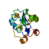
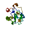
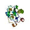
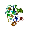
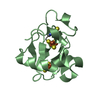
- PDB-6fd1: 7-FE FERREDOXIN FROM AZOTOBACTER VINELANDII LOW TEMPERATURE, 1.35 A -
+ Open data

- Basic information
| Entry | Database: PDB / ID: 6fd1 | ||||||
|---|---|---|---|---|---|---|---|
| Title | 7-FE FERREDOXIN FROM AZOTOBACTER VINELANDII LOW TEMPERATURE, 1.35 A | ||||||
 Components Components | 7-FE FERREDOXIN I (FD1) | ||||||
 Keywords Keywords | ELECTRON TRANSPORT / IRON-SULFUR | ||||||
| Function / homology |  Function and homology information Function and homology information3 iron, 4 sulfur cluster binding / 4 iron, 4 sulfur cluster binding / electron transfer activity / DNA binding / metal ion binding Similarity search - Function | ||||||
| Biological species |  Azotobacter vinelandii (bacteria) Azotobacter vinelandii (bacteria) | ||||||
| Method |  X-RAY DIFFRACTION / SYNCHROTRON / MOLECULAR REPLACEMENT / Resolution: 1.35 Å X-RAY DIFFRACTION / SYNCHROTRON / MOLECULAR REPLACEMENT / Resolution: 1.35 Å | ||||||
 Authors Authors | Stout, C.D. / Stura, E.A. / Mcree, D.E. | ||||||
 Citation Citation | Journal: J.Mol.Biol. / Year: 1998 Title: Structure of Azotobacter vinelandii 7Fe ferredoxin at 1.35 A resolution and determination of the [Fe-S] bonds with 0.01 A accuracy. Authors: Stout, C.D. / Stura, E.A. / McRee, D.E. | ||||||
| History |
|
- Structure visualization
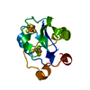
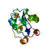
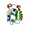
| Structure viewer | Molecule: MolmilJmol/JSmol |
|---|
- Downloads & links
-Download
| PDBx/mmCIF format | 6fd1.cif.gz | 90.2 KB | Display | PDBx/mmCIF format |
|---|---|---|---|---|
| PDB format | pdb6fd1.ent.gz | 67.3 KB | Display | PDB format |
| PDBx/mmJSON format | 6fd1.json.gz | Tree view | PDBx/mmJSON format | |
| Others |  Other downloads Other downloads |
-Validation report
| Arichive directory | https://data.pdbj.org/pub/pdb/validation_reports/fd/6fd1ftp://data.pdbj.org/pub/pdb/validation_reports/fd/6fd1 | HTTPS FTP |
|---|
-Related structure data
| Related structure data | 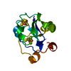 5fd1S S: Starting model for refinement |
|---|---|
| Similar structure data |


-Links
 PDBj
PDBj


- Assembly
| Deposited unit | 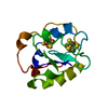
| ||||||||
|---|---|---|---|---|---|---|---|---|---|
| 1 |
| ||||||||
| Unit cell |
|
-Components
| #1: Protein | Mass: 12059.530 Da / Num. of mol.: 1 / Source method: isolated from a natural source / Source: (natural) Azotobacter vinelandii (bacteria) / References: UniProt: P00214 |
|---|---|
| #2: Chemical | ChemComp-SF4 /   Mass: 351.640 Da / Num. of mol.: 1 / Source method: obtained synthetically / Formula: Fe4S4 Mass: 351.640 Da / Num. of mol.: 1 / Source method: obtained synthetically / Formula: Fe4S4 |
| #3: Chemical | ChemComp-F3S /   Mass: 295.795 Da / Num. of mol.: 1 / Source method: obtained synthetically / Formula: Fe3S4 Mass: 295.795 Da / Num. of mol.: 1 / Source method: obtained synthetically / Formula: Fe3S4 |
| #4: Water | ChemComp-HOH /  Mass: 18.015 Da / Num. of mol.: 162 / Source method: isolated from a natural source / Formula: H2O Mass: 18.015 Da / Num. of mol.: 162 / Source method: isolated from a natural source / Formula: H2O |
-Experimental details
-Experiment
| Experiment | Method: X-RAY DIFFRACTION / Number of used crystals: 1 |
|---|
- Sample preparation
| Crystal | Density Matthews: 2.89 Å3/Da / Density % sol: 57.38 % Description: ALL DATA INCLUDING NEGATIVE I'S WERE USED WERE PROCESSED IN POINT GROUP 422 - PRESERVE BIJVOET PAIRS | ||||||||||||||||||||
|---|---|---|---|---|---|---|---|---|---|---|---|---|---|---|---|---|---|---|---|---|---|
| Crystal grow | pH: 7.8 Details: PROTEIN CRYSTALLIZED FROM 4.8M AMMONIUM SULFATE, 0.45M TRIS-HCL, PH 7.8 | ||||||||||||||||||||
| Crystal grow | *PLUS Method: batch methodDetails: anaerobic conditions, Martin, A.E., (1990) Proc. Natl. Acad. Sci. USA., 87, 598. | ||||||||||||||||||||
| Components of the solutions | *PLUS
|
-Data collection
| Diffraction | Mean temperature: 100 K |
|---|---|
| Diffraction source | Source: SYNCHROTRON / Site: SSRL  / Beamline: BL7-1 / Wavelength: 1.08 / Beamline: BL7-1 / Wavelength: 1.08 |
| Detector | Type: MARRESEARCH / Detector: IMAGE PLATE / Date: Apr 1, 1997 |
| Radiation | Monochromatic (M) / Laue (L): M / Scattering type: x-ray |
| Radiation wavelength | Wavelength: 1.08 Å / Relative weight: 1 |
| Reflection | Resolution: 1.35→100 Å / Num. obs: 57315 / % possible obs: 96.5 % / Redundancy: 3.3 % / Rsym value: 0.057 / Net I/σ(I): 21.3 |
| Reflection shell | Resolution: 1.35→1.38 Å / Redundancy: 2.4 % / Mean I/σ(I) obs: 1.7 / Rsym value: 0.747 / % possible all: 72 |
| Reflection | *PLUS Lowest resolution: 50 Å / Rmerge(I) obs: 0.057 |
- Processing
| Software |
| |||||||||||||||||||||||||||||||||
|---|---|---|---|---|---|---|---|---|---|---|---|---|---|---|---|---|---|---|---|---|---|---|---|---|---|---|---|---|---|---|---|---|---|---|
| Refinement | Method to determine structure: MOLECULAR REPLACEMENT Starting model: PDB ENTRY 5FD1 Resolution: 1.35→100 Å / Num. parameters: 9211 / Num. restraintsaints: 10844 / Cross valid method: R-FREE / σ(F): 0 / StereochEM target val spec case: FE CLUSTERS NOT RESTRAINED / Stereochemistry target values: ENGH & HUBER Details: FREE R USED UNTIL LAST CYCLE WHEN ALL DATA WAS INCLUDED.
| |||||||||||||||||||||||||||||||||
| Solvent computation | Solvent model: MOEWS & KRETSINGER | |||||||||||||||||||||||||||||||||
| Refine analyze | Num. disordered residues: 1 / Occupancy sum hydrogen: 776 / Occupancy sum non hydrogen: 1011.5 | |||||||||||||||||||||||||||||||||
| Refinement step | Cycle: LAST / Resolution: 1.35→100 Å
| |||||||||||||||||||||||||||||||||
| Refine LS restraints |
| |||||||||||||||||||||||||||||||||
| Software | *PLUS Name: SHELXL-97 / Classification: refinement | |||||||||||||||||||||||||||||||||
| Refinement | *PLUS Lowest resolution: 9999 Å / Rfactor all: 0.15 | |||||||||||||||||||||||||||||||||
| Solvent computation | *PLUS | |||||||||||||||||||||||||||||||||
| Displacement parameters | *PLUS |
