 Movie
Movie Controller


+ Open data

- Basic information
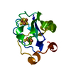
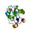
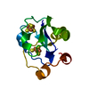
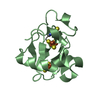
| Entry | Database: PDB / ID: 1g6b | ||||||
|---|---|---|---|---|---|---|---|
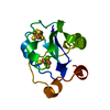
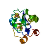
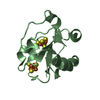
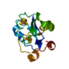
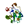
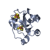
| Title | CRYSTAL STRUCTURE OF P47S MUTANT OF FERREDOXIN I | ||||||
 Components Components | 7FE FERREDOXIN I | ||||||
 Keywords Keywords | ELECTRON TRANSPORT / Iron-sulfur clusters / ferredoxin | ||||||
| Function / homology |  Function and homology information Function and homology information3 iron, 4 sulfur cluster binding / 4 iron, 4 sulfur cluster binding / electron transfer activity / DNA binding / metal ion binding Similarity search - Function | ||||||
| Biological species |  Azotobacter vinelandii (bacteria) Azotobacter vinelandii (bacteria) | ||||||
| Method |  X-RAY DIFFRACTION / MOLECULAR REPLACEMENT / Resolution: 1.9 Å X-RAY DIFFRACTION / MOLECULAR REPLACEMENT / Resolution: 1.9 Å | ||||||
 Authors Authors | Stout, C.D. / Burgess, B.K. / Bonagura, C.A. / Jung, Y.S. | ||||||
 Citation Citation | Journal: J.Biol.Chem. / Year: 2002 Title: Azotobacter vinelandii ferredoxin I: a sequence and structure comparison approach to alteration of [4Fe-4S]2+/+ reduction potential. Authors: Chen, K. / Jung, Y.S. / Bonagura, C.A. / Tilley, G.J. / Prasad, G.S. / Sridhar, V. / Armstrong, F.A. / Stout, C.D. / Burgess, B.K. | ||||||
| History |
|
- Structure visualization
| Structure viewer | Molecule: MolmilJmol/JSmol |
|---|
- Downloads & links
-Download
| PDBx/mmCIF format | 1g6b.cif.gz | 40.1 KB | Display | PDBx/mmCIF format |
|---|---|---|---|---|
| PDB format | pdb1g6b.ent.gz | 26.3 KB | Display | PDB format |
| PDBx/mmJSON format | 1g6b.json.gz | Tree view | PDBx/mmJSON format | |
| Others |  Other downloads Other downloads |
-Validation report
| Arichive directory | https://data.pdbj.org/pub/pdb/validation_reports/g6/1g6bftp://data.pdbj.org/pub/pdb/validation_reports/g6/1g6b | HTTPS FTP |
|---|
-Related structure data
| Related structure data | 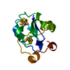 1g3oC 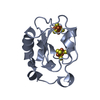 1gaoC 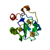 7fd1S S: Starting model for refinement C: citing same article ( |
|---|---|
| Similar structure data |

-Links
 PDBj
PDBj


- Assembly
| Deposited unit | 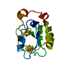
| ||||||||
|---|---|---|---|---|---|---|---|---|---|
| 1 |
| ||||||||
| Unit cell |
|
-Components
| #1: Protein | Mass: 12049.493 Da / Num. of mol.: 1 / Source method: isolated from a natural source / Details: MUTANT DESIGNED TO TEST REDUCTION POTENTIAL SHIFT / Source: (natural) Azotobacter vinelandii (bacteria) / References: UniProt: P00214 |
|---|---|
| #2: Chemical | ChemComp-SF4 /   Mass: 351.640 Da / Num. of mol.: 1 / Source method: obtained synthetically / Formula: Fe4S4 Mass: 351.640 Da / Num. of mol.: 1 / Source method: obtained synthetically / Formula: Fe4S4 |
| #3: Chemical | ChemComp-F3S /   Mass: 295.795 Da / Num. of mol.: 1 / Source method: obtained synthetically / Formula: Fe3S4 Mass: 295.795 Da / Num. of mol.: 1 / Source method: obtained synthetically / Formula: Fe3S4 |
| #4: Water | ChemComp-HOH /  Mass: 18.015 Da / Num. of mol.: 215 / Source method: isolated from a natural source / Formula: H2O Mass: 18.015 Da / Num. of mol.: 215 / Source method: isolated from a natural source / Formula: H2O |
-Experimental details
-Experiment
| Experiment | Method: X-RAY DIFFRACTION / Number of used crystals: 1 |
|---|
- Sample preparation
| Crystal | Density Matthews: 2.89 Å3/Da / Density % sol: 57.37 % | |||||||||||||||||||||||||
|---|---|---|---|---|---|---|---|---|---|---|---|---|---|---|---|---|---|---|---|---|---|---|---|---|---|---|
| Crystal grow | Temperature: 277 K / Method: vapor diffusion / pH: 7.5 Details: Ammonium sulfate, tris buffer, pH 7.5, VAPOR DIFFUSION, temperature 277.0K | |||||||||||||||||||||||||
| Crystal grow | *PLUS Temperature: 2 ℃ / pH: 7.4 / Details: Shen, B., (1994) J. Biol. Chem., 269, 8564. | |||||||||||||||||||||||||
| Components of the solutions | *PLUS
|
-Data collection
| Diffraction | Mean temperature: 100 K |
|---|---|
| Diffraction source | Source: ROTATING ANODE / Type: RIGAKU RU200 / Wavelength: 1.5418 Å |
| Detector | Type: RIGAKU RAXIS / Detector: IMAGE PLATE / Date: Jan 1, 2000 |
| Radiation | Protocol: SINGLE WAVELENGTH / Monochromatic (M) / Laue (L): M / Scattering type: x-ray |
| Radiation wavelength | Wavelength: 1.5418 Å / Relative weight: 1 |
| Reflection | Resolution: 1.9→24 Å / Num. all: 83321 / Num. obs: 11740 / % possible obs: 99.9 % / Observed criterion σ(F): 0 / Observed criterion σ(I): 0 / Redundancy: 7.1 % / Biso Wilson estimate: 17.8 Å2 / Rmerge(I) obs: 0.085 / Net I/σ(I): 21.8 |
| Reflection shell | Resolution: 1.9→1.93 Å / Redundancy: 7 % / Rmerge(I) obs: 0.392 / Mean I/σ(I) obs: 3.7 / Num. unique all: 557 / % possible all: 99.9 |
| Reflection | *PLUS Num. measured all: 83321 |
- Processing
| Software |
| |||||||||||||||||||||||||
|---|---|---|---|---|---|---|---|---|---|---|---|---|---|---|---|---|---|---|---|---|---|---|---|---|---|---|
| Refinement | Method to determine structure: MOLECULAR REPLACEMENT Starting model: 7FD1 Resolution: 1.9→24 Å / Isotropic thermal model: Isotropic / σ(F): 0 / σ(I): 0 / Stereochemistry target values: Xplor3.8
| |||||||||||||||||||||||||
| Refinement step | Cycle: LAST / Resolution: 1.9→24 Å
| |||||||||||||||||||||||||
| Refine LS restraints |
| |||||||||||||||||||||||||
| Software | *PLUS Name: X-PLOR / Version: 3.851 / Classification: refinement | |||||||||||||||||||||||||
| Refinement | *PLUS Lowest resolution: 24 Å / σ(F): 0 / % reflection Rfree: 5 % / Rfactor obs: 0.24 / Rfactor Rwork: 0.24 | |||||||||||||||||||||||||
| Solvent computation | *PLUS | |||||||||||||||||||||||||
| Displacement parameters | *PLUS | |||||||||||||||||||||||||
| Refine LS restraints | *PLUS
|
