 Movie
Movie Controller


+ Open data

- Basic information
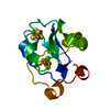
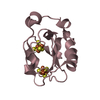
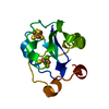
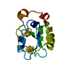
| Entry | Database: PDB / ID: 1axq | ||||||
|---|---|---|---|---|---|---|---|
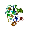
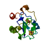
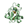
| Title | FERRICYANIDE OXIDIZED FDI | ||||||
 Components Components | FERREDOXIN | ||||||
 Keywords Keywords | ELECTRON TRANSPORT | ||||||
| Function / homology |  Function and homology information Function and homology information3 iron, 4 sulfur cluster binding / 4 iron, 4 sulfur cluster binding / electron transfer activity / DNA binding / metal ion binding Similarity search - Function | ||||||
| Biological species |  Azotobacter vinelandii (bacteria) Azotobacter vinelandii (bacteria) | ||||||
| Method |  X-RAY DIFFRACTION / MOLECULAR REPLACEMENT / Resolution: 2.1 Å X-RAY DIFFRACTION / MOLECULAR REPLACEMENT / Resolution: 2.1 Å | ||||||
 Authors Authors | Stout, C.D. | ||||||
 Citation Citation | Journal: J.Biol.Inorg.Chem. / Year: 1998 Title: Crystal structures of ferricyanide-oxidized [Fe-S] clusters in Azotobacter vinelandii ferredoxin I. Authors: Sridhar, V. / Prasad, G.S. / Burgess, B.K. / Stout, C.D. | ||||||
| History |
|
- Structure visualization
| Structure viewer | Molecule: MolmilJmol/JSmol |
|---|
- Downloads & links
-Download
| PDBx/mmCIF format | 1axq.cif.gz | 33.8 KB | Display | PDBx/mmCIF format |
|---|---|---|---|---|
| PDB format | pdb1axq.ent.gz | 22.4 KB | Display | PDB format |
| PDBx/mmJSON format | 1axq.json.gz | Tree view | PDBx/mmJSON format | |
| Others |  Other downloads Other downloads |
-Validation report
| Arichive directory | https://data.pdbj.org/pub/pdb/validation_reports/ax/1axqftp://data.pdbj.org/pub/pdb/validation_reports/ax/1axq | HTTPS FTP |
|---|
-Related structure data
| Related structure data | 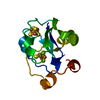 5fd1S S: Starting model for refinement |
|---|---|
| Similar structure data |


-Links
 PDBj
PDBj


- Assembly
| Deposited unit | 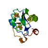
| ||||||||
|---|---|---|---|---|---|---|---|---|---|
| 1 |
| ||||||||
| Unit cell |
|
-Components
| #1: Protein | Mass: 12059.530 Da / Num. of mol.: 1 Source method: isolated from a genetically manipulated source Details: THIS 7FE FERREDOXIN FROM A. VINELANDII TREATED WITH 9\:1 MOLE RATIO OF FERRICYANIDE IN THE CRYSTALS. THE [FE-S] CLUSTERS HAVE BEEN PARTIALLY OXIDIZED. Source: (gene. exp.) Azotobacter vinelandii (bacteria) / Strain: JG100Description: WILD TYPE FERREDOXIN OVER-EXPRESSED FROM A PLASMID IN A. VINELANDII Gene: FDXA / Plasmid: PKT320 / Gene (production host): FDXA / Production host: Azotobacter vinelandii (bacteria) / Strain (production host): JG100 / References: UniProt: P00214 |
|---|---|
| #2: Chemical | ChemComp-SF4 /   Mass: 351.640 Da / Num. of mol.: 1 / Source method: obtained synthetically / Formula: Fe4S4 Mass: 351.640 Da / Num. of mol.: 1 / Source method: obtained synthetically / Formula: Fe4S4 |
| #3: Chemical | ChemComp-F3S /   Mass: 295.795 Da / Num. of mol.: 1 / Source method: obtained synthetically / Formula: Fe3S4 Mass: 295.795 Da / Num. of mol.: 1 / Source method: obtained synthetically / Formula: Fe3S4 |
-Experimental details
-Experiment
| Experiment | Method: X-RAY DIFFRACTION / Number of used crystals: 1 |
|---|
- Sample preparation
| Crystal | Density Matthews: 2.8 Å3/Da / Density % sol: 59.9 % | |||||||||||||||||||||||||
|---|---|---|---|---|---|---|---|---|---|---|---|---|---|---|---|---|---|---|---|---|---|---|---|---|---|---|
| Crystal grow | pH: 7.8 / Details: 4.8 M AMMONIUM SULFATE, 0.45 M TRIS-HCL PH 7.8 | |||||||||||||||||||||||||
| Crystal grow | *PLUS Temperature: 2 ℃ / pH: 7.4 / Method: vapor diffusion / Details: Stout, C.D., (1979) J. Biol. Chem., 254, 3598. | |||||||||||||||||||||||||
| Components of the solutions | *PLUS
|
-Data collection
| Diffraction | Mean temperature: 291 K |
|---|---|
| Diffraction source | Source: ROTATING ANODE / Type: RIGAKU RUH2R / Wavelength: 1.5418 |
| Detector | Type: SIEMENS / Detector: AREA DETECTOR / Date: Jan 10, 1996 |
| Radiation | Monochromator: GRAPHITE(002) / Monochromatic (M) / Laue (L): M / Scattering type: x-ray |
| Radiation wavelength | Wavelength: 1.5418 Å / Relative weight: 1 |
| Reflection | Resolution: 2.1→20 Å / Num. obs: 8124 / % possible obs: 88.4 % / Observed criterion σ(I): 0 / Redundancy: 3.9 % / Rsym value: 0.101 / Net I/σ(I): 11.2 |
| Reflection shell | Resolution: 2.1→2.25 Å / Redundancy: 3.9 % / Mean I/σ(I) obs: 1.5 / % possible all: 88.4 |
| Reflection | *PLUS Rmerge(I) obs: 0.101 |
- Processing
| Software |
| ||||||||||||||||||||||||||||||||||||||||||||||||||||||||||||
|---|---|---|---|---|---|---|---|---|---|---|---|---|---|---|---|---|---|---|---|---|---|---|---|---|---|---|---|---|---|---|---|---|---|---|---|---|---|---|---|---|---|---|---|---|---|---|---|---|---|---|---|---|---|---|---|---|---|---|---|---|---|
| Refinement | Method to determine structure: MOLECULAR REPLACEMENT Starting model: PDB ENTRY 5FD1 Resolution: 2.1→8 Å / Data cutoff high absF: 10000000 / Data cutoff low absF: 0.001 / σ(F): 0 Details: MODIFIED PARAMETER AND TOPOLOGY FILES TO REFINE OXIDIZED, DISTORTED [FE-S] CLUSTERS OXIDIZED [FE-S] CLUSTERS WERE REFINED WITH MODIFIED TOPOLOGY AND PARAMETER FILES TO ALLOW FIT OF FE AND S ...Details: MODIFIED PARAMETER AND TOPOLOGY FILES TO REFINE OXIDIZED, DISTORTED [FE-S] CLUSTERS OXIDIZED [FE-S] CLUSTERS WERE REFINED WITH MODIFIED TOPOLOGY AND PARAMETER FILES TO ALLOW FIT OF FE AND S TO DENSITY RESULTING FOR FERRICYANIDE OXIDATION OF THE CRYSTALS. THE RESULTING GEOMETRY IS DISTORTED COMPARED TO IDEAL [FE-S] CLUSTERS.
| ||||||||||||||||||||||||||||||||||||||||||||||||||||||||||||
| Displacement parameters | Biso mean: 19.3 Å2 | ||||||||||||||||||||||||||||||||||||||||||||||||||||||||||||
| Refinement step | Cycle: LAST / Resolution: 2.1→8 Å
| ||||||||||||||||||||||||||||||||||||||||||||||||||||||||||||
| Refine LS restraints |
| ||||||||||||||||||||||||||||||||||||||||||||||||||||||||||||
| Software | *PLUS Name: X-PLOR / Version: 3.8 / Classification: refinement | ||||||||||||||||||||||||||||||||||||||||||||||||||||||||||||
| Refine LS restraints | *PLUS
|
