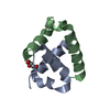
- PDB-6nid: Crystal structure of a human calcium/calmodulin dependent serine ... -
+
Open data
ID or keywords:
Loading...
-
Basic information
Entry
Database: PDB / ID: 6nid
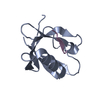
Title
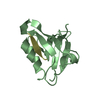
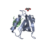
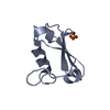
Crystal structure of a human calcium/calmodulin dependent serine protein kinase (CASK) PDZ domain in complex with Neurexin-1 peptide
Components
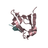
Neurexin-1
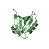
Peripheral plasma membrane protein CASK
Keywords
protein binding/peptide / PDZ domain / MAGUK protein family / peripheral plasma membrane protein / protein binding / c-terminal peptide binding / Neurexin / protein binding-peptide complex
Function / homology
Function and homology information
negative regulation of cellular response to growth factor stimulus / neuroligin clustering involved in postsynaptic membrane assembly / gephyrin clustering involved in postsynaptic density assembly / postsynaptic density protein 95 clustering / postsynaptic membrane assembly / positive regulation of synapse maturation / neuroligin family protein binding / vocal learning / neurexin family protein binding / GMP kinase activity ...negative regulation of cellular response to growth factor stimulus / neuroligin clustering involved in postsynaptic membrane assembly / gephyrin clustering involved in postsynaptic density assembly / postsynaptic density protein 95 clustering / postsynaptic membrane assembly / positive regulation of synapse maturation / neuroligin family protein binding / vocal learning / neurexin family protein binding / GMP kinase activity / negative regulation of wound healing / neuron cell-cell adhesion / Dopamine Neurotransmitter Release Cycle / regulation of neurotransmitter secretion / nuclear lamina / calcium ion import / vocalization behavior / acetylcholine receptor binding / neurotransmitter secretion / Nephrin family interactions / positive regulation of synapse assembly / Assembly and cell surface presentation of NMDA receptors / Sensory processing of sound by outer hair cells of the cochlea / Neurexins and neuroligins / Sensory processing of sound by inner hair cells of the cochlea / neuromuscular process controlling balance / Syndecan interactions / ciliary membrane / positive regulation of calcium ion import / regulation of synaptic vesicle exocytosis / adult behavior / Non-integrin membrane-ECM interactions / basement membrane / negative regulation of cell-matrix adhesion / negative regulation of keratinocyte proliferation / social behavior / positive regulation of synaptic transmission, glutamatergic / synapse assembly / cell adhesion molecule binding / establishment of localization in cell / axon guidance / positive regulation of excitatory postsynaptic potential / learning / calcium channel regulator activity / Schaffer collateral - CA1 synapse / nuclear matrix / cell-cell junction / intracellular protein localization / actin cytoskeleton / signaling receptor activity / presynaptic membrane / nuclear membrane / vesicle / basolateral plasma membrane / chemical synaptic transmission / calmodulin binding / non-specific serine/threonine protein kinase / cell adhesion / signaling receptor binding / protein serine kinase activity / focal adhesion / protein serine/threonine kinase activity / neuronal cell body / calcium ion binding / nucleolus / cell surface / endoplasmic reticulum / positive regulation of transcription by RNA polymerase II / ATP binding / plasma membrane / cytoplasm / cytosol Similarity search - Function
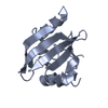
PeripheralplasmamembraneproteinCASK / hCASK / Calcium/calmodulin-dependent serine protein kinase / Protein lin-2 homolog
Mass: 10118.886 Da / Num. of mol.: 3 Source method: isolated from a genetically manipulated source Details: M 485 Expression artifact G 486 Expression artifact Source: (gene. exp.) Homo sapiens (human) / Gene: CASK, LIN2 / Production host: Escherichia coli (E. coli) References: UniProt: O14936, non-specific serine/threonine protein kinase
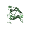
#2: Protein/peptide
Neurexin-1 / Neurexin I-alpha / Neurexin-1-alpha
Mass: 1318.517 Da / Num. of mol.: 3 / Source method: obtained synthetically / Source: (synth.) Homo sapiens (human) / References: UniProt: Q9ULB1
In the structure databanks used in Yorodumi, some data are registered as the other names, "COVID-19 virus" and "2019-nCoV". Here are the details of the virus and the list of structure data.
Jan 31, 2019. EMDB accession codes are about to change! (news from PDBe EMDB page)
EMDB accession codes are about to change! (news from PDBe EMDB page)
The allocation of 4 digits for EMDB accession codes will soon come to an end. Whilst these codes will remain in use, new EMDB accession codes will include an additional digit and will expand incrementally as the available range of codes is exhausted. The current 4-digit format prefixed with “EMD-” (i.e. EMD-XXXX) will advance to a 5-digit format (i.e. EMD-XXXXX), and so on. It is currently estimated that the 4-digit codes will be depleted around Spring 2019, at which point the 5-digit format will come into force.
The EM Navigator/Yorodumi systems omit the EMD- prefix.
Related info.:Q: What is EMD? / ID/Accession-code notation in Yorodumi/EM Navigator
Yorodumi is a browser for structure data from EMDB, PDB, SASBDB, etc.
This page is also the successor to EM Navigator detail page, and also detail information page/front-end page for Omokage search.
The word "yorodu" (or yorozu) is an old Japanese word meaning "ten thousand". "mi" (miru) is to see.
Related info.:EMDB / PDB / SASBDB / Comparison of 3 databanks / Yorodumi Search / Aug 31, 2016. New EM Navigator & Yorodumi / Yorodumi Papers / Jmol/JSmol / Function and homology information / Changes in new EM Navigator and Yorodumi
 Movie
Movie Controller

 Yorodumi
 Open data
 Basic information
 Components
 Keywords
 Function and homology information
 Homo sapiens (human)
 X-RAY DIFFRACTION /
  Authors
 United States, 1items
  Citation
 Structure visualization
 Downloads & links
 Other downloads
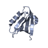


 PDBj
PDBj


 Assembly
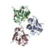


 Mass: 62.068 Da / Num. of mol.: 1 / Source method: isolated from a natural source / Formula: C2H6O2
 Mass: 18.015 Da / Num. of mol.: 211 / Source method: isolated from a natural source / Formula: H2O
 Sample preparation
 Processing