positive regulation of protein folding / negative regulation of protein refolding / Regulation of HSF1-mediated heat shock response / dATP binding / Hsp70 protein binding / response to bacterium / : / protein-folding chaperone binding / protein dimerization activity / protein domain specific binding ...positive regulation of protein folding / negative regulation of protein refolding / Regulation of HSF1-mediated heat shock response / dATP binding / Hsp70 protein binding / response to bacterium / : / protein-folding chaperone binding / protein dimerization activity / protein domain specific binding / protein-containing complex binding / protein-containing complex / identical protein binding / cytosol 類似検索 - 分子機能
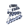
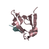
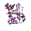
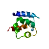
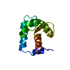
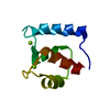
Single alpha-helices involved in coiled-coils or other helix-helix interfaces - #3420 / Hsp70-interacting protein, N-terminal / Hsp70-interacting protein N N-terminal domain / STI1/HOP, DP domain / STI1/HOP, DP domain / Heat shock chaperonin-binding / Heat shock chaperonin-binding motif. / Single alpha-helices involved in coiled-coils or other helix-helix interfaces / Tetratricopeptide repeat / TPR repeat region circular profile. ...Single alpha-helices involved in coiled-coils or other helix-helix interfaces - #3420 / Hsp70-interacting protein, N-terminal / Hsp70-interacting protein N N-terminal domain / STI1/HOP, DP domain / STI1/HOP, DP domain / Heat shock chaperonin-binding / Heat shock chaperonin-binding motif. / Single alpha-helices involved in coiled-coils or other helix-helix interfaces / Tetratricopeptide repeat / TPR repeat region circular profile. / TPR repeat profile. / Tetratricopeptide repeats / Tetratricopeptide repeat / Helix non-globular / Special / Tetratricopeptide-like helical domain superfamily 類似検索 - ドメイン・相同性
プロトコル: SINGLE WAVELENGTH / 単色(M)・ラウエ(L): M / 散乱光タイプ: x-ray
放射波長
波長: 0.90004 Å / 相対比: 1
Reflection
冗長度: 7.3 % / Av σ(I) over netI: 4.6 / 数: 94893 / Rmerge(I) obs: 0.112 / Rsym value: 0.112 / D res high: 1.493 Å / D res low: 43.895 Å / Num. obs: 12978 / % possible obs: 98.8
 ムービー
ムービー コントローラー


 データを開く
 基本情報
基本情報 要素
要素 キーワード
キーワード 機能・相同性情報
 X線回折 /
  データ登録者
 引用
引用 構造の表示
構造の表示 ダウンロードとリンク
 その他のダウンロード
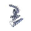


 PDBj
PDBj


 集合体
集合体


 分子量: 92.094 Da / 分子数: 1 / 由来タイプ: 合成 / 式: C3H8O3
 分子量: 18.015 Da / 分子数: 147 / 由来タイプ: 天然 / 式: H2O
 試料調製
試料調製 / ビームライン: ID29 / 波長: 0.90004 Å
 解析
解析