 Movie
Movie Controller


+ Open data

- Basic information
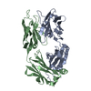
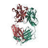
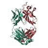
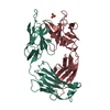
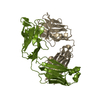
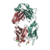
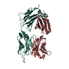
| Entry | Database: PDB / ID: 1rfd | ||||||
|---|---|---|---|---|---|---|---|
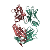
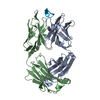
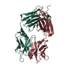
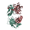
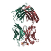
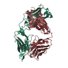
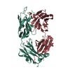
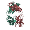
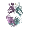
| Title | ANTI-COCAINE ANTIBODY M82G2 | ||||||
 Components Components |
| ||||||
 Keywords Keywords | IMMUNE SYSTEM / ANTI-COCAINE ANTIBODY / FAB | ||||||
| Function / homology | Immunoglobulins / Immunoglobulin-like / Sandwich / Mainly Beta Function and homology information Function and homology information | ||||||
| Biological species |  | ||||||
| Method |  X-RAY DIFFRACTION / SYNCHROTRON / MOLECULAR REPLACEMENT / Resolution: 2.09 Å X-RAY DIFFRACTION / SYNCHROTRON / MOLECULAR REPLACEMENT / Resolution: 2.09 Å | ||||||
 Authors Authors | Pozharski, E. / Hewagama, A. / Shanafelt, A.B. / Petsko, G.A. / Ringe, D. | ||||||
 Citation Citation | Journal: J.Mol.Biol. / Year: 2005 Title: Diversity in hapten recognition: structural study of an anti-cocaine antibody M82G2. Authors: Pozharski, E. / Moulin, A. / Hewagama, A. / Shanafelt, A.B. / Petsko, G.A. / Ringe, D. | ||||||
| History |
| ||||||
| Remark 999 | SEQUENCE NO DATABASE REFERENCE SEQUENCE WAS FOUND FOR EITHER PROTEIN IN THIS STRUCTURE AT THE TIME ...SEQUENCE NO DATABASE REFERENCE SEQUENCE WAS FOUND FOR EITHER PROTEIN IN THIS STRUCTURE AT THE TIME OF PROCESSING. |
- Structure visualization
| Structure viewer | Molecule: MolmilJmol/JSmol |
|---|
- Downloads & links
-Download
| PDBx/mmCIF format | 1rfd.cif.gz | 100.8 KB | Display | PDBx/mmCIF format |
|---|---|---|---|---|
| PDB format | pdb1rfd.ent.gz | 76 KB | Display | PDB format |
| PDBx/mmJSON format | 1rfd.json.gz | Tree view | PDBx/mmJSON format | |
| Others |  Other downloads Other downloads |
-Validation report
| Summary document | 1rfd_validation.pdf.gz | 431.9 KB | Display | wwPDB validaton report |
|---|---|---|---|---|
| Full document | 1rfd_full_validation.pdf.gz | 438.2 KB | Display | |
| Data in XML | 1rfd_validation.xml.gz | 20.1 KB | Display | |
| Data in CIF | 1rfd_validation.cif.gz | 29 KB | Display | |
| Arichive directory | https://data.pdbj.org/pub/pdb/validation_reports/rf/1rfdftp://data.pdbj.org/pub/pdb/validation_reports/rf/1rfd | HTTPS FTP |
-Related structure data
| Related structure data |  1q72C 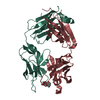 1qygSC S: Starting model for refinement C: citing same article ( |
|---|---|
| Similar structure data |


-Links
 PDBj
PDBj

- Assembly
| Deposited unit | 
| ||||||||
|---|---|---|---|---|---|---|---|---|---|
| 1 |
| ||||||||
| Unit cell |
| ||||||||
| Components on special symmetry positions |
|
-Components
| #1: Antibody | Mass: 23884.365 Da / Num. of mol.: 1 / Source method: isolated from a natural source / Source: (natural) |
|---|---|
| #2: Antibody | Mass: 23519.264 Da / Num. of mol.: 1 / Source method: isolated from a natural source / Source: (natural) |
| #3: Water | ChemComp-HOH /  Mass: 18.015 Da / Num. of mol.: 237 / Source method: isolated from a natural source / Formula: H2O Mass: 18.015 Da / Num. of mol.: 237 / Source method: isolated from a natural source / Formula: H2O |
| Has protein modification | Y |
-Experimental details
-Experiment
| Experiment | Method: X-RAY DIFFRACTION / Number of used crystals: 1 |
|---|
- Sample preparation
| Crystal | Density Matthews: 2.58 Å3/Da / Density % sol: 52.39 % |
|---|---|
| Crystal grow | Temperature: 298 K / Method: vapor diffusion, hanging drop / pH: 5.2 Details: 2M AMMONIUM SULFATE, 5% ISO-PROPANOL, pH 5.20, VAPOR DIFFUSION, HANGING DROP, temperature 298K |
-Data collection
| Diffraction | Mean temperature: 110 K |
|---|---|
| Diffraction source | Source: SYNCHROTRON / Site: SSRL  / Beamline: BL7-1 / Wavelength: 1.08 Å / Beamline: BL7-1 / Wavelength: 1.08 Å |
| Detector | Type: MARRESEARCH / Detector: IMAGE PLATE / Date: Nov 30, 2002 |
| Radiation | Protocol: SINGLE WAVELENGTH / Monochromatic (M) / Laue (L): M / Scattering type: x-ray |
| Radiation wavelength | Wavelength: 1.08 Å / Relative weight: 1 |
| Reflection | Resolution: 2.09→30 Å / Num. all: 26932 / Num. obs: 26932 / % possible obs: 90.6 % / Observed criterion σ(F): 0 / Observed criterion σ(I): 0 / Redundancy: 6.2 % / Biso Wilson estimate: 38.5 Å2 / Rmerge(I) obs: 0.067 / Net I/σ(I): 25.4 |
| Reflection shell | Resolution: 2.09→2.16 Å / Redundancy: 5.8 % / Mean I/σ(I) obs: 4.8 / Num. unique all: 2603 / % possible all: 88.9 |
- Processing
| Software |
| ||||||||||||||||||||||||||||||||||||
|---|---|---|---|---|---|---|---|---|---|---|---|---|---|---|---|---|---|---|---|---|---|---|---|---|---|---|---|---|---|---|---|---|---|---|---|---|---|
| Refinement | Method to determine structure: MOLECULAR REPLACEMENT Starting model: PDB ENTRY 1QYG Resolution: 2.09→30 Å / Isotropic thermal model: isotropic / Cross valid method: THROUGHOUT / σ(F): 0 / σ(I): 0 / Stereochemistry target values: Engh & Huber Details: BULK SOLVENT MASK CORRECTED TO EXCLUDE INTERNAL CAVITIES
| ||||||||||||||||||||||||||||||||||||
| Displacement parameters | Biso mean: 0.303 Å2
| ||||||||||||||||||||||||||||||||||||
| Refine analyze |
| ||||||||||||||||||||||||||||||||||||
| Refinement step | Cycle: LAST / Resolution: 2.09→30 Å
| ||||||||||||||||||||||||||||||||||||
| Refine LS restraints |
| ||||||||||||||||||||||||||||||||||||
| LS refinement shell | Resolution: 2.09→2.16 Å
|
