 Movie
Movie Controller

[English] 日本語
 Yorodumi
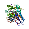
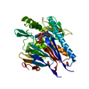
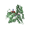
- PDB-1qbg: CRYSTAL STRUCTURE OF HUMAN DT-DIAPHORASE (NAD(P)H OXIDOREDUCTASE) -
+ Open data

- Basic information
| Entry | Database: PDB / ID: 1qbg | ||||||
|---|---|---|---|---|---|---|---|
| Title | CRYSTAL STRUCTURE OF HUMAN DT-DIAPHORASE (NAD(P)H OXIDOREDUCTASE) | ||||||
 Components Components | NAD(P)H DEHYDROGENASE [QUINONE] 1 | ||||||
 Keywords Keywords | OXIDOREDUCTASE / QUINONE / FAD / DT-DIAPHORASE | ||||||
| Function / homology |  Function and homology information Function and homology informationubiquinone metabolic process / vitamin E metabolic process / NAD(P)H dehydrogenase (quinone) / NADPH dehydrogenase (quinone) activity / cytochrome-b5 reductase activity, acting on NAD(P)H / vitamin K metabolic process / NADH dehydrogenase (quinone) (non-electrogenic) activity / NAD(P)H dehydrogenase (quinone) activity / NFE2L2 regulating anti-oxidant/detoxification enzymes / Regulation of ornithine decarboxylase (ODC) ...ubiquinone metabolic process / vitamin E metabolic process / NAD(P)H dehydrogenase (quinone) / NADPH dehydrogenase (quinone) activity / cytochrome-b5 reductase activity, acting on NAD(P)H / vitamin K metabolic process / NADH dehydrogenase (quinone) (non-electrogenic) activity / NAD(P)H dehydrogenase (quinone) activity / NFE2L2 regulating anti-oxidant/detoxification enzymes / Regulation of ornithine decarboxylase (ODC) / synaptic transmission, cholinergic / negative regulation of ferroptosis / nitric oxide biosynthetic process / xenobiotic metabolic process / removal of superoxide radicals / cell redox homeostasis / protein catabolic process / negative regulation of protein catabolic process / response to toxic substance / protein polyubiquitination / cellular response to oxidative stress / response to oxidative stress / response to lipopolysaccharide / innate immune response / synapse / RNA binding / identical protein binding / nucleus / cytosol / cytoplasm Similarity search - Function | ||||||
| Biological species |  Homo sapiens (human) Homo sapiens (human) | ||||||
| Method |  X-RAY DIFFRACTION / Resolution: 2.3 Å X-RAY DIFFRACTION / Resolution: 2.3 Å | ||||||
 Authors Authors | Skelly, J.V. / Sanderson, M.R. / Suter, D.A. / Baumann, U. / Gregory, D.S. / Bennett, M. / Hobbs, S.M. / Neidle, S. | ||||||
 Citation Citation | Journal: J.Med.Chem. / Year: 1999 Title: Crystal structure of human DT-diaphorase: a model for interaction with the cytotoxic prodrug 5-(aziridin-1-yl)-2,4-dinitrobenzamide (CB1954). Authors: Skelly, J.V. / Sanderson, M.R. / Suter, D.A. / Baumann, U. / Read, M.A. / Gregory, D.S. / Bennett, M. / Hobbs, S.M. / Neidle, S. | ||||||
| History |
|
- Structure visualization
| Structure viewer | Molecule: MolmilJmol/JSmol |
|---|
- Downloads & links
-Download
| PDBx/mmCIF format | 1qbg.cif.gz | 222.9 KB | Display | PDBx/mmCIF format |
|---|---|---|---|---|
| PDB format | pdb1qbg.ent.gz | 181.1 KB | Display | PDB format |
| PDBx/mmJSON format | 1qbg.json.gz | Tree view | PDBx/mmJSON format | |
| Others |  Other downloads Other downloads |
-Validation report
| Arichive directory | https://data.pdbj.org/pub/pdb/validation_reports/qb/1qbgftp://data.pdbj.org/pub/pdb/validation_reports/qb/1qbg | HTTPS FTP |
|---|
-Related structure data
| Similar structure data |
|---|


-Links
 PDBj
PDBj
- Assembly
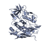
| Deposited unit | 
| ||||||||||
|---|---|---|---|---|---|---|---|---|---|---|---|
| 1 | 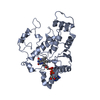
| ||||||||||
| 2 | 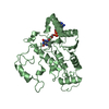
| ||||||||||
| 3 | 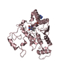
| ||||||||||
| 4 | 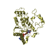
| ||||||||||
| 5 | 
| ||||||||||
| 6 | 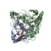
| ||||||||||
| Unit cell |
|
-Components
| #1: Protein | Mass: 30719.365 Da / Num. of mol.: 4 Source method: isolated from a genetically manipulated source Source: (gene. exp.) Homo sapiens (human) / Production host:  #2: Chemical | ChemComp-FAD /   Mass: 785.550 Da / Num. of mol.: 4 / Source method: obtained synthetically / Formula: C27H33N9O15P2 / Comment: FAD*YM Mass: 785.550 Da / Num. of mol.: 4 / Source method: obtained synthetically / Formula: C27H33N9O15P2 / Comment: FAD*YM |
|---|
-Experimental details
-Experiment
| Experiment | Method: X-RAY DIFFRACTION / Number of used crystals: 1 |
|---|
- Sample preparation
| Crystal | Density Matthews: 2.44 Å3/Da / Density % sol: 49.66 % | ||||||||||||||||||||
|---|---|---|---|---|---|---|---|---|---|---|---|---|---|---|---|---|---|---|---|---|---|
| Crystal grow | Temperature: 291 K / Method: vapor diffusion, sitting drop / pH: 7.5 Details: Sitting drop, PEG4000, CYMAL-3 detergent, sodium phosphate, pH 7.5, VAPOR DIFFUSION, SITTING DROP, temperature 291K | ||||||||||||||||||||
| Crystal | *PLUS Density % sol: 47 % | ||||||||||||||||||||
| Crystal grow | *PLUS Temperature: 18 ℃ | ||||||||||||||||||||
| Components of the solutions | *PLUS
|
-Data collection
| Diffraction | Mean temperature: 298 K |
|---|---|
| Diffraction source | Source: ROTATING ANODE / Type: RIGAKU RU200 / Wavelength: 1.5418 |
| Detector | Type: RIGAKU RAXIS II / Detector: IMAGE PLATE / Date: Oct 15, 1996 |
| Radiation | Protocol: SINGLE WAVELENGTH / Monochromatic (M) / Laue (L): M / Scattering type: x-ray |
| Radiation wavelength | Wavelength: 1.5418 Å / Relative weight: 1 |
| Reflection | Resolution: 2.3→40 Å / Num. all: 47496 / Num. obs: 43746 / Observed criterion σ(F): 2 / Observed criterion σ(I): 4 / Rmerge(I) obs: 0.132 / Net I/σ(I): 4.98 |
| Reflection shell | Resolution: 2.3→2.4 Å / Rmerge(I) obs: 0.6 / Num. unique all: 4439 / % possible all: 70 |
| Reflection | *PLUS % possible obs: 81.5 % / Num. measured all: 193332 |
- Processing
| Software |
| ||||||||||||||||||||
|---|---|---|---|---|---|---|---|---|---|---|---|---|---|---|---|---|---|---|---|---|---|
| Refinement | Resolution: 2.3→40 Å / Cross valid method: THROUGHOUT / σ(F): 4 / σ(I): 2 / Stereochemistry target values: Engh & Huber / Details: used x-plor procedure
| ||||||||||||||||||||
| Refinement step | Cycle: LAST / Resolution: 2.3→40 Å
| ||||||||||||||||||||
| Software | *PLUS Name: X-PLOR / Version: 3.851 / Classification: refinement | ||||||||||||||||||||
| Refinement | *PLUS Highest resolution: 2.3 Å / Lowest resolution: 40 Å / % reflection Rfree: 4.3 % / Rfactor Rfree: 0.28 / Rfactor Rwork: 0.232 | ||||||||||||||||||||
| Solvent computation | *PLUS | ||||||||||||||||||||
| Displacement parameters | *PLUS |
