 Movie
Movie Controller

[English] 日本語
 Yorodumi
- PDB-1f06: THREE DIMENSIONAL STRUCTURE OF THE TERNARY COMPLEX OF CORYNEBACTE... -
+ Open data

- Basic information
| Entry | Database: PDB / ID: 1f06 | ||||||
|---|---|---|---|---|---|---|---|
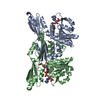
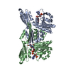
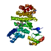
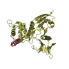
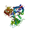
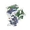
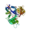
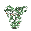
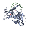
| Title | THREE DIMENSIONAL STRUCTURE OF THE TERNARY COMPLEX OF CORYNEBACTERIUM GLUTAMICUM DIAMINOPIMELATE DEHYDROGENASE NADPH-L-2-AMINO-6-METHYLENE-PIMELATE | ||||||
 Components Components | MESO-DIAMINOPIMELATE D-DEHYDROGENASE | ||||||
 Keywords Keywords | OXIDOREDUCTASE / Enzyme-NADPH-inhibitor ternary complex | ||||||
| Function / homology |  Function and homology information Function and homology informationdiaminopimelate dehydrogenase / diaminopimelate dehydrogenase activity / : / : Similarity search - Function | ||||||
| Biological species |  Corynebacterium glutamicum (bacteria) Corynebacterium glutamicum (bacteria) | ||||||
| Method |  X-RAY DIFFRACTION / Resolution: 2.1 Å X-RAY DIFFRACTION / Resolution: 2.1 Å | ||||||
 Authors Authors | Cirilli, M. / Scapin, G. / Sutherland, A. / Caplan, J.F. / Vederas, J.C. / Blanchard, J.S. | ||||||
 Citation Citation | Journal: Protein Sci. / Year: 2000 Title: The three-dimensional structure of the ternary complex of Corynebacterium glutamicum diaminopimelate dehydrogenase-NADPH-L-2-amino-6-methylene-pimelate. Authors: Cirilli, M. / Scapin, G. / Sutherland, A. / Vederas, J.C. / Blanchard, J.S. | ||||||
| History |
|
- Structure visualization
| Structure viewer | Molecule: MolmilJmol/JSmol |
|---|
- Downloads & links
-Download
| PDBx/mmCIF format | 1f06.cif.gz | 136.9 KB | Display | PDBx/mmCIF format |
|---|---|---|---|---|
| PDB format | pdb1f06.ent.gz | 107.4 KB | Display | PDB format |
| PDBx/mmJSON format | 1f06.json.gz | Tree view | PDBx/mmJSON format | |
| Others |  Other downloads Other downloads |
-Validation report
| Arichive directory | https://data.pdbj.org/pub/pdb/validation_reports/f0/1f06ftp://data.pdbj.org/pub/pdb/validation_reports/f0/1f06 | HTTPS FTP |
|---|
-Related structure data
| Related structure data | |
|---|---|
| Similar structure data |


-Links
 PDBj
PDBj

- Assembly
| Deposited unit | 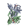
| ||||||||
|---|---|---|---|---|---|---|---|---|---|
| 1 | 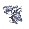
| ||||||||
| 2 | 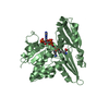
| ||||||||
| 3 |
| ||||||||
| Unit cell |
|
-Components
| #1: Protein | Mass: 35242.328 Da / Num. of mol.: 2 Source method: isolated from a genetically manipulated source Source: (gene. exp.) Corynebacterium glutamicum (bacteria) / Plasmid: PET23B / Production host: #2: Chemical |   Mass: 745.421 Da / Num. of mol.: 2 / Source method: obtained synthetically / Formula: C21H30N7O17P3 Mass: 745.421 Da / Num. of mol.: 2 / Source method: obtained synthetically / Formula: C21H30N7O17P3#3: Chemical | ChemComp-2NP / |   Mass: 187.193 Da / Num. of mol.: 1 / Source method: obtained synthetically / Formula: C8H13NO4 Mass: 187.193 Da / Num. of mol.: 1 / Source method: obtained synthetically / Formula: C8H13NO4#4: Water | ChemComp-HOH / |  Mass: 18.015 Da / Num. of mol.: 116 / Source method: isolated from a natural source / Formula: H2O Mass: 18.015 Da / Num. of mol.: 116 / Source method: isolated from a natural source / Formula: H2O |
|---|
-Experimental details
-Experiment
| Experiment | Method: X-RAY DIFFRACTION / Number of used crystals: 1 |
|---|
- Sample preparation
| Crystal | Density Matthews: 2.84 Å3/Da / Density % sol: 56.68 % | ||||||||||||||||||||||||||||||||||||||||
|---|---|---|---|---|---|---|---|---|---|---|---|---|---|---|---|---|---|---|---|---|---|---|---|---|---|---|---|---|---|---|---|---|---|---|---|---|---|---|---|---|---|
| Crystal grow | Temperature: 298 K / Method: vapor diffusion, hanging drop / pH: 6.8 Details: PEG 8000, magnesium acetate, cacodylate, pH 6.8, VAPOR DIFFUSION, HANGING DROP, temperature 298.0K | ||||||||||||||||||||||||||||||||||||||||
| Crystal grow | *PLUS pH: 7.5 | ||||||||||||||||||||||||||||||||||||||||
| Components of the solutions | *PLUS
|
-Data collection
| Diffraction | Mean temperature: 298 K |
|---|---|
| Diffraction source | Source: ROTATING ANODE / Type: RIGAKU RU300 / Wavelength: 1.5418 |
| Detector | Type: XENTRONICS / Detector: AREA DETECTOR / Date: Jul 28, 1999 |
| Radiation | Protocol: SINGLE WAVELENGTH / Monochromatic (M) / Laue (L): M / Scattering type: x-ray |
| Radiation wavelength | Wavelength: 1.5418 Å / Relative weight: 1 |
| Reflection | Resolution: 2.1→48.6 Å / Num. all: 111792 / Num. obs: 111416 / % possible obs: 86.9 % / Observed criterion σ(F): 2 / Observed criterion σ(I): 2 / Redundancy: 2.82 % / Biso Wilson estimate: 32.7 Å2 / Rmerge(I) obs: 0.076 / Net I/σ(I): 8.615 |
| Reflection shell | Resolution: 2.11→2.25 Å / Redundancy: 1.5 % / Rmerge(I) obs: 0.224 / % possible all: 47.3 |
| Reflection | *PLUS Num. obs: 39441 |
| Reflection shell | *PLUS % possible obs: 67 % |
- Processing
| Software |
| ||||||||||||||||||||
|---|---|---|---|---|---|---|---|---|---|---|---|---|---|---|---|---|---|---|---|---|---|
| Refinement | Resolution: 2.1→48.6 Å / σ(F): 2 / σ(I): 4 / Stereochemistry target values: Engh & Huber
| ||||||||||||||||||||
| Refinement step | Cycle: LAST / Resolution: 2.1→48.6 Å
| ||||||||||||||||||||
| Refine LS restraints |
| ||||||||||||||||||||
| Software | *PLUS Name: X-PLOR / Version: 3.851 / Classification: refinement | ||||||||||||||||||||
| Refinement | *PLUS Highest resolution: 2.1 Å / Lowest resolution: 48.6 Å / σ(F): 2 / Rfactor obs: 0.178 | ||||||||||||||||||||
| Solvent computation | *PLUS | ||||||||||||||||||||
| Displacement parameters | *PLUS | ||||||||||||||||||||
| Refine LS restraints | *PLUS
|
