 Movie
Movie Controller

[English] 日本語
 Yorodumi
- PDB-3dap: C. GLUTAMICUM DAP DEHYDROGENASE IN COMPLEX WITH NADP+ AND THE INH... -
+ Open data

- Basic information
| Entry | Database: PDB / ID: 3dap | ||||||
|---|---|---|---|---|---|---|---|
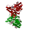
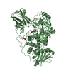
| Title | C. GLUTAMICUM DAP DEHYDROGENASE IN COMPLEX WITH NADP+ AND THE INHIBITOR 5S-ISOXAZOLINE | ||||||
 Components Components | DIAMINOPIMELIC ACID DEHYDROGENASE | ||||||
 Keywords Keywords | OXIDOREDUCTASE / NADP / DEHYDROGENASE / D-AMINO ACID DEHYDROGENASE / LYSINE BIOSYNTHESIS / ASYMMETRIC DIMER / INHIBITOR | ||||||
| Function / homology |  Function and homology information Function and homology informationdiaminopimelate dehydrogenase / diaminopimelate dehydrogenase activity / : / : Similarity search - Function | ||||||
| Biological species |  Corynebacterium glutamicum (bacteria) Corynebacterium glutamicum (bacteria) | ||||||
| Method |  X-RAY DIFFRACTION / Resolution: 2.2 Å X-RAY DIFFRACTION / Resolution: 2.2 Å | ||||||
 Authors Authors | Scapin, G. / Cirilli, M. / Reddy, S.G. / Gao, Y. / Vederas, J.C. / Blanchard, J.S. | ||||||
 Citation Citation | Journal: Biochemistry / Year: 1998 Title: Substrate and inhibitor binding sites in Corynebacterium glutamicum diaminopimelate dehydrogenase. Authors: Scapin, G. / Cirilli, M. / Reddy, S.G. / Gao, Y. / Vederas, J.C. / Blanchard, J.S. #1: Journal: Biochemistry / Year: 1996Title: Three-Dimensional Structure of Meso-Diaminopimelic Acid Dehydrogenase from Corynebacterium Glutamicum Authors: Scapin, G. / Reddy, S.G. / Blanchard, J.S. #2: Journal: Proteins / Year: 1996Title: Expression, Purification, and Crystallization of Meso-Diaminopimelate Dehydrogenase from Corynebacterium Glutamicum Authors: Reddy, S.G. / Scapin, G. / Blanchard, J.S. | ||||||
| History |
|
- Structure visualization
| Structure viewer | Molecule: MolmilJmol/JSmol |
|---|
- Downloads & links
-Download
| PDBx/mmCIF format | 3dap.cif.gz | 137.4 KB | Display | PDBx/mmCIF format |
|---|---|---|---|---|
| PDB format | pdb3dap.ent.gz | 108.1 KB | Display | PDB format |
| PDBx/mmJSON format | 3dap.json.gz | Tree view | PDBx/mmJSON format | |
| Others |  Other downloads Other downloads |
-Validation report
| Arichive directory | https://data.pdbj.org/pub/pdb/validation_reports/da/3dapftp://data.pdbj.org/pub/pdb/validation_reports/da/3dap | HTTPS FTP |
|---|
-Related structure data


-Links
 PDBj
PDBj

- Assembly
| Deposited unit | 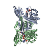
| ||||||||||||
|---|---|---|---|---|---|---|---|---|---|---|---|---|---|
| 1 |
| ||||||||||||
| Unit cell |
| ||||||||||||
| Noncrystallographic symmetry (NCS) | NCS oper:
|
-Components
| #1: Protein | Mass: 35242.328 Da / Num. of mol.: 2 Source method: isolated from a genetically manipulated source Source: (gene. exp.) Corynebacterium glutamicum (bacteria) / Cell line: BL21 / Gene: DAPDH / Plasmid: PET23A / Species (production host): Escherichia coli / Gene (production host): DAPDH / Production host: #2: Chemical |   Mass: 745.421 Da / Num. of mol.: 2 / Source method: obtained synthetically / Formula: C21H30N7O17P3 Mass: 745.421 Da / Num. of mol.: 2 / Source method: obtained synthetically / Formula: C21H30N7O17P3#3: Chemical | ChemComp-DA3 / ( |   Mass: 202.165 Da / Num. of mol.: 1 / Source method: obtained synthetically / Formula: C7H10N2O5 Mass: 202.165 Da / Num. of mol.: 1 / Source method: obtained synthetically / Formula: C7H10N2O5#4: Water | ChemComp-HOH / |  Mass: 18.015 Da / Num. of mol.: 133 / Source method: isolated from a natural source / Formula: H2O Mass: 18.015 Da / Num. of mol.: 133 / Source method: isolated from a natural source / Formula: H2O |
|---|
-Experimental details
-Experiment
| Experiment | Method: X-RAY DIFFRACTION / Number of used crystals: 1 |
|---|
- Sample preparation
| Crystal | Density Matthews: 2.8 Å3/Da / Density % sol: 65 % | ||||||||||||||||||||||||||||||||||||||||||||||||||
|---|---|---|---|---|---|---|---|---|---|---|---|---|---|---|---|---|---|---|---|---|---|---|---|---|---|---|---|---|---|---|---|---|---|---|---|---|---|---|---|---|---|---|---|---|---|---|---|---|---|---|---|
| Crystal grow | pH: 6.5 Details: 13-17% PEG 8000 IN 100 MM NA-CACODYLATE, PH 6.5, 150-300 MM MG-ACETATE CRYSTAL | ||||||||||||||||||||||||||||||||||||||||||||||||||
| Crystal | *PLUS Density % sol: 60 % | ||||||||||||||||||||||||||||||||||||||||||||||||||
| Crystal grow | *PLUS Method: vapor diffusion, hanging drop | ||||||||||||||||||||||||||||||||||||||||||||||||||
| Components of the solutions | *PLUS
|
-Data collection
| Diffraction | Mean temperature: 290 K |
|---|---|
| Diffraction source | Source: ROTATING ANODE / Type: RIGAKU RUH2R / Wavelength: 1.5418 |
| Detector | Type: SIEMENS / Detector: AREA DETECTOR / Date: Jun 1, 1997 |
| Radiation | Monochromator: NI FILTER / Monochromatic (M) / Laue (L): M / Scattering type: x-ray |
| Radiation wavelength | Wavelength: 1.5418 Å / Relative weight: 1 |
| Reflection | Resolution: 2→19 Å / Num. obs: 46303 / % possible obs: 88.9 % / Redundancy: 2 % / Biso Wilson estimate: 19.9 Å2 / Rsym value: 0.074 / Net I/σ(I): 10 |
| Reflection shell | Resolution: 2→2.1 Å / Redundancy: 1.5 % / Mean I/σ(I) obs: 2.5 / Rsym value: 0.209 / % possible all: 77.7 |
| Reflection | *PLUS Num. measured all: 70458 / Rmerge(I) obs: 0.074 |
| Reflection shell | *PLUS % possible obs: 77.7 % / Num. unique obs: 4342 / Num. measured obs: 5440 / Rmerge(I) obs: 0.209 |
- Processing
| Software |
| ||||||||||||||||||||||||||||||||||||||||||||||||||||||||||||
|---|---|---|---|---|---|---|---|---|---|---|---|---|---|---|---|---|---|---|---|---|---|---|---|---|---|---|---|---|---|---|---|---|---|---|---|---|---|---|---|---|---|---|---|---|---|---|---|---|---|---|---|---|---|---|---|---|---|---|---|---|---|
| Refinement | Resolution: 2.2→19 Å / Data cutoff high absF: 1000000 / Data cutoff low absF: 0.001 / Cross valid method: THROUGHOUT / σ(F): 2
| ||||||||||||||||||||||||||||||||||||||||||||||||||||||||||||
| Displacement parameters | Biso mean: 24.5 Å2 | ||||||||||||||||||||||||||||||||||||||||||||||||||||||||||||
| Refinement step | Cycle: LAST / Resolution: 2.2→19 Å
| ||||||||||||||||||||||||||||||||||||||||||||||||||||||||||||
| Refine LS restraints |
| ||||||||||||||||||||||||||||||||||||||||||||||||||||||||||||
| LS refinement shell | Resolution: 2.2→2.3 Å / Total num. of bins used: 8
| ||||||||||||||||||||||||||||||||||||||||||||||||||||||||||||
| Xplor file |
| ||||||||||||||||||||||||||||||||||||||||||||||||||||||||||||
| Software | *PLUS Name: X-PLOR / Version: 3.851 / Classification: refinement | ||||||||||||||||||||||||||||||||||||||||||||||||||||||||||||
| Refinement | *PLUS Rfactor all: 0.179 | ||||||||||||||||||||||||||||||||||||||||||||||||||||||||||||
| Solvent computation | *PLUS | ||||||||||||||||||||||||||||||||||||||||||||||||||||||||||||
| Displacement parameters | *PLUS | ||||||||||||||||||||||||||||||||||||||||||||||||||||||||||||
| Refine LS restraints | *PLUS
|
