SEQUENCE THE CONSTRUCT WAS EXPRESSED WITH A PURIFICATION TAG MGSDKIHHHHHHENLYFQG. THE TAG WAS ...SEQUENCE THE CONSTRUCT WAS EXPRESSED WITH A PURIFICATION TAG MGSDKIHHHHHHENLYFQG. THE TAG WAS REMOVED WITH TEV PROTEASE LEAVING ONLY A GLYCINE (0) FOLLOWED BY THE TARGET SEQUENCE.
Resolution: 2→69.5 Å / Num. obs: 53921 / % possible obs: 91.6 % / Redundancy: 3.2 % / Rmerge(I) obs: 0.067 / Net I/σ(I): 9.53
Reflection shell
Diffraction-ID: 1
Resolution (Å)
% possible obs (%)
Rmerge(I) obs
Mean I/σ(I) obs
Num. measured obs
Num. unique obs
% possible all
2-2.07
79.6
0.424
2.05
10419
8517
79.6
2.07-2.15
85
0.599
2.73
10995
8992
2.15-2.25
87.7
0.599
3.42
12002
9794
2.25-2.37
90.5
0.599
4.37
12170
9971
2.37-2.52
91.4
0.599
5.39
12343
10138
2.52-2.71
93.1
0.599
6.81
12114
9964
2.71-2.99
94.2
0.599
9.6
12711
10484
2.99-3.42
96.7
0.599
14.04
21791
10512
3.42-4.3
98.4
0.599
17.82
33399
10680
4.3-69.5
98.7
0.599
25.04
33563
10852
-
Phasing
Phasing
Method: MAD
-
Processing
Software
Name
Version
Classification
NB
REFMAC
5.2.0005
refinement
XSCALE
datascaling
PDB_EXTRACT
1.601
dataextraction
XDS
datareduction
SHELX
phasing
SHARP
phasing
Refinement
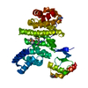
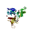
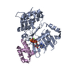
Method to determine structure: MAD / Resolution: 2→69.5 Å / Cor.coef. Fo:Fc: 0.965 / Cor.coef. Fo:Fc free: 0.94 / SU B: 8.233 / SU ML: 0.124 / TLS residual ADP flag: LIKELY RESIDUAL / Cross valid method: THROUGHOUT / ESU R: 0.172 / ESU R Free: 0.161 Stereochemistry target values: MAXIMUM LIKELIHOOD WITH PHASES Details: 1. HYDROGENS HAVE BEEN ADDED IN THE RIDING POSITIONS 2. A MET-INHIBITION PROTOCOL WAS USED FOR SELENOMETHIONINE INCORPORATION DURING PROTEIN EXPRESSION. THE OCCUPANCY OF THE SE ATOMS IN THE ...Details: 1. HYDROGENS HAVE BEEN ADDED IN THE RIDING POSITIONS 2. A MET-INHIBITION PROTOCOL WAS USED FOR SELENOMETHIONINE INCORPORATION DURING PROTEIN EXPRESSION. THE OCCUPANCY OF THE SE ATOMS IN THE MSE RESIDUES WAS REDUCED TO 0.75 TO ACCOUNT FOR THE REDUCED SCATTERING POWER DUE TO PARTIAL S-MET INCORPORATION. 3. ADP AND MG WERE MODELED BASED ON ELECTRON DENSITY AND THE PROTEIN'S PROPOSED FUNCTION AS AN ATPASE. 4. RESIDUES 115-119 ARE DISORDERED IN EACH CHAIN AND WERE NOT MODELED.
Rfactor
Num. reflection
% reflection
Selection details
Rfree
0.226
2733
5.1 %
RANDOM
Rwork
0.174
-
-
-
all
0.177
-
-
-
obs
0.17656
51161
97.51 %
-
Solvent computation
Ion probe radii: 0.8 Å / Shrinkage radii: 0.8 Å / VDW probe radii: 1.2 Å / Solvent model: MASK
Displacement parameters
Biso mean: 36.569 Å2
Baniso -1
Baniso -2
Baniso -3
1-
-2.3 Å2
0 Å2
-0.71 Å2
2-
-
1.17 Å2
0 Å2
3-
-
-
0.88 Å2
Refinement step
Cycle: LAST / Resolution: 2→69.5 Å
Protein
Nucleic acid
Ligand
Solvent
Total
Num. atoms
5688
0
84
347
6119
Refine LS restraints
Refine-ID
Type
Dev ideal
Dev ideal target
Number
X-RAY DIFFRACTION
r_bond_refined_d
0.014
0.022
5942
X-RAY DIFFRACTION
r_bond_other_d
0.001
0.02
5552
X-RAY DIFFRACTION
r_angle_refined_deg
1.425
2
8020
X-RAY DIFFRACTION
r_angle_other_deg
0.818
3
12865
X-RAY DIFFRACTION
r_dihedral_angle_1_deg
5.772
5
720
X-RAY DIFFRACTION
r_dihedral_angle_2_deg
33.827
23.791
277
X-RAY DIFFRACTION
r_dihedral_angle_3_deg
14.787
15
1092
X-RAY DIFFRACTION
r_dihedral_angle_4_deg
17.074
15
43
X-RAY DIFFRACTION
r_chiral_restr
0.082
0.2
884
X-RAY DIFFRACTION
r_gen_planes_refined
0.005
0.02
6483
X-RAY DIFFRACTION
r_gen_planes_other
0.001
0.02
1256
X-RAY DIFFRACTION
r_nbd_refined
0.214
0.2
1193
X-RAY DIFFRACTION
r_nbd_other
0.176
0.2
5483
X-RAY DIFFRACTION
r_nbtor_refined
0.185
0.2
2880
X-RAY DIFFRACTION
r_nbtor_other
0.082
0.2
3445
X-RAY DIFFRACTION
r_xyhbond_nbd_refined
0.163
0.2
321
X-RAY DIFFRACTION
r_xyhbond_nbd_other
0.018
0.2
1
X-RAY DIFFRACTION
r_symmetry_vdw_refined
0.119
0.2
7
X-RAY DIFFRACTION
r_symmetry_vdw_other
0.218
0.2
53
X-RAY DIFFRACTION
r_symmetry_hbond_refined
0.12
0.2
12
X-RAY DIFFRACTION
r_mcbond_it
1.961
3
3629
X-RAY DIFFRACTION
r_mcbond_other
0.485
3
1440
X-RAY DIFFRACTION
r_mcangle_it
2.923
5
5686
X-RAY DIFFRACTION
r_scbond_it
4.934
8
2634
X-RAY DIFFRACTION
r_scangle_it
6.568
11
2324
LS refinement shell
Resolution: 2→2.052 Å / Total num. of bins used: 20
In the structure databanks used in Yorodumi, some data are registered as the other names, "COVID-19 virus" and "2019-nCoV". Here are the details of the virus and the list of structure data.
Jan 31, 2019. EMDB accession codes are about to change! (news from PDBe EMDB page)
EMDB accession codes are about to change! (news from PDBe EMDB page)
The allocation of 4 digits for EMDB accession codes will soon come to an end. Whilst these codes will remain in use, new EMDB accession codes will include an additional digit and will expand incrementally as the available range of codes is exhausted. The current 4-digit format prefixed with “EMD-” (i.e. EMD-XXXX) will advance to a 5-digit format (i.e. EMD-XXXXX), and so on. It is currently estimated that the 4-digit codes will be depleted around Spring 2019, at which point the 5-digit format will come into force.
The EM Navigator/Yorodumi systems omit the EMD- prefix.
Related info.:Q: What is EMD? / ID/Accession-code notation in Yorodumi/EM Navigator
Yorodumi is a browser for structure data from EMDB, PDB, SASBDB, etc.
This page is also the successor to EM Navigator detail page, and also detail information page/front-end page for Omokage search.
The word "yorodu" (or yorozu) is an old Japanese word meaning "ten thousand". "mi" (miru) is to see.
Related info.:EMDB / PDB / SASBDB / Comparison of 3 databanks / Yorodumi Search / Aug 31, 2016. New EM Navigator & Yorodumi / Yorodumi Papers / Jmol/JSmol / Function and homology information / Changes in new EM Navigator and Yorodumi
 Movie
Movie Controller

 Yorodumi
 Open data
 Basic information
 Components
 Keywords
 Function and homology information
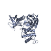
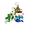
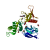
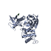
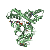
 Sulfolobus solfataricus (archaea)
 X-RAY DIFFRACTION /
  Authors
 Citation
 Structure visualization
 Downloads & links
 Other downloads
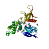


 PDBj
PDBj Assembly


 Mass: 24.305 Da / Num. of mol.: 2 / Source method: obtained synthetically / Formula: Mg
 Mass: 427.201 Da / Num. of mol.: 2 / Source method: obtained synthetically / Formula: C10H15N5O10P2 / Comment: ADP, energy-carrying molecule*YM
 Mass: 62.068 Da / Num. of mol.: 7 / Source method: obtained synthetically / Formula: C2H6O2
 Mass: 18.015 Da / Num. of mol.: 347 / Source method: isolated from a natural source / Formula: H2O
 Sample preparation
 / Beamline: 23-ID-D / Wavelength: 0.95372, 0.979996
 Processing