 Movie
Movie Controller

[English] 日本語
 Yorodumi
- PDB-1muj: Crystal structure of murine class II MHC I-Ab in complex with a h... -
+ Open data

- Basic information
| Entry | Database: PDB / ID: 1muj | ||||||
|---|---|---|---|---|---|---|---|
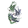
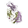
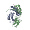
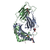
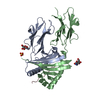
| Title | Crystal structure of murine class II MHC I-Ab in complex with a human CLIP peptide | ||||||
 Components Components |
| ||||||
 Keywords Keywords | IMMUNE SYSTEM / I-AB / CLIP / COMPLEX / ANTIGEN | ||||||
| Function / homology |  Function and homology information Function and homology informationpositive regulation of antigen processing and presentation / positive regulation of alpha-beta T cell activation / negative regulation of peptide secretion / macrophage migration inhibitory factor signaling pathway / NOS2-CD74 complex / MHC class II protein binding, via antigen binding groove / antigen processing and presentation of endogenous antigen / positive regulation of dendritic cell antigen processing and presentation / negative regulation of T cell differentiation / positive regulation of macrophage migration inhibitory factor signaling pathway ...positive regulation of antigen processing and presentation / positive regulation of alpha-beta T cell activation / negative regulation of peptide secretion / macrophage migration inhibitory factor signaling pathway / NOS2-CD74 complex / MHC class II protein binding, via antigen binding groove / antigen processing and presentation of endogenous antigen / positive regulation of dendritic cell antigen processing and presentation / negative regulation of T cell differentiation / positive regulation of macrophage migration inhibitory factor signaling pathway / macrophage migration inhibitory factor receptor complex / protein trimerization / macrophage migration inhibitory factor binding / positive regulation of cytokine-mediated signaling pathway / positive regulation of T-helper 1 type immune response / T cell activation involved in immune response / T cell selection / positive regulation of type 2 immune response / positive regulation of prostaglandin biosynthetic process / host-mediated suppression of symbiont invasion / MHC class II protein binding / B cell affinity maturation / negative thymic T cell selection / negative regulation of mature B cell apoptotic process / protein antigen binding / positive regulation of monocyte differentiation / positive thymic T cell selection / CD4 receptor binding / prostaglandin biosynthetic process / vacuole / positive regulation of chemokine (C-X-C motif) ligand 2 production / cytokine receptor activity / positive regulation of neutrophil chemotaxis / positive regulation of macrophage cytokine production / positive regulation of T cell differentiation / antigen processing and presentation / negative regulation of intrinsic apoptotic signaling pathway in response to DNA damage by p53 class mediator / transport vesicle membrane / regulation of macrophage activation / negative regulation of DNA damage response, signal transduction by p53 class mediator / cytokine binding / immunoglobulin mediated immune response / nitric-oxide synthase binding / response to type II interferon / toxic substance binding / positive regulation of chemokine production / multivesicular body / positive regulation of B cell proliferation / MHC class II antigen presentation / protein folding chaperone / lysosomal lumen / negative regulation of cell migration / Cell surface interactions at the vascular wall / trans-Golgi network membrane / positive regulation of interleukin-8 production / Developmental Lineage of Pancreatic Ductal Cells / lumenal side of endoplasmic reticulum membrane / ER to Golgi transport vesicle membrane / clathrin-coated endocytic vesicle membrane / intracellular protein transport / MHC class II protein complex / positive regulation of interleukin-6 production / cellular response to type II interferon / antigen processing and presentation of exogenous peptide antigen via MHC class II / peptide antigen binding / positive regulation of fibroblast proliferation / endocytic vesicle membrane / MHC class II protein complex binding / late endosome / amyloid-beta binding / protein-containing complex assembly / adaptive immune response / early endosome / positive regulation of ERK1 and ERK2 cascade / positive regulation of canonical NF-kappaB signal transduction / lysosome / protein stabilization / Golgi membrane / external side of plasma membrane / lysosomal membrane / ubiquitin protein ligase binding / positive regulation of gene expression / negative regulation of apoptotic process / cell surface / Golgi apparatus / protein-containing complex / extracellular exosome / membrane / identical protein binding / nucleus / plasma membrane / cytoplasm Similarity search - Function | ||||||
| Biological species |   Homo sapiens (human) Homo sapiens (human) | ||||||
| Method |  X-RAY DIFFRACTION / SYNCHROTRON / MOLECULAR REPLACEMENT / Resolution: 2.15 Å X-RAY DIFFRACTION / SYNCHROTRON / MOLECULAR REPLACEMENT / Resolution: 2.15 Å | ||||||
 Authors Authors | Zhu, Y. / Wilson, I.A. | ||||||
 Citation Citation | Journal: J.Mol.Biol. / Year: 2003 Title: Crystal structure of MHC class II I-Ab in complex with a human CLIP peptide: Prediction of an I-Ab peptide-binding motif Authors: Zhu, Y. / Rudensky, A.Y. / Teyton, A.L. / Wilson, I.A. | ||||||
| History |
|
- Structure visualization
| Structure viewer | Molecule: MolmilJmol/JSmol |
|---|
- Downloads & links
-Download
| PDBx/mmCIF format | 1muj.cif.gz | 96.7 KB | Display | PDBx/mmCIF format |
|---|---|---|---|---|
| PDB format | pdb1muj.ent.gz | 72.8 KB | Display | PDB format |
| PDBx/mmJSON format | 1muj.json.gz | Tree view | PDBx/mmJSON format | |
| Others |  Other downloads Other downloads |
-Validation report
| Arichive directory | https://data.pdbj.org/pub/pdb/validation_reports/mu/1mujftp://data.pdbj.org/pub/pdb/validation_reports/mu/1muj | HTTPS FTP |
|---|
-Related structure data
| Similar structure data |
|---|


-Links
 PDBj
PDBj


- Assembly
| Deposited unit | 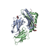
| ||||||||
|---|---|---|---|---|---|---|---|---|---|
| 1 |
| ||||||||
| Unit cell |
|
-Components
| #1: Protein | Mass: 21428.828 Da / Num. of mol.: 1 Fragment: EXTRACELLULAR ALPHA-1 and EXTRACELLULAR ALPHA-2 domains Source method: isolated from a genetically manipulated source Source: (gene. exp.)  | ||||
|---|---|---|---|---|---|
| #2: Protein | Mass: 23192.846 Da / Num. of mol.: 1 Fragment: EXTRACELLULAR BETA-1 and EXTRACELLULAR BETA-2 domains Source method: isolated from a genetically manipulated source Source: (gene. exp.) | ||||
| #3: Protein/peptide | Mass: 3696.436 Da / Num. of mol.: 1 Source method: isolated from a genetically manipulated source Details: The peptide was covalently linked to the N-terminus of chain B. Source: (gene. exp.) Homo sapiens (human) / Cell line (production host): S2 / Production host: | ||||
| #4: Sugar |   Type: D-saccharide, beta linking / Mass: 221.208 Da / Num. of mol.: 2 Type: D-saccharide, beta linking / Mass: 221.208 Da / Num. of mol.: 2Source method: isolated from a genetically manipulated source Formula: C8H15NO6 #5: Water | ChemComp-HOH / |  Mass: 18.015 Da / Num. of mol.: 171 / Source method: isolated from a natural source / Formula: H2O Mass: 18.015 Da / Num. of mol.: 171 / Source method: isolated from a natural source / Formula: H2OHas protein modification | Y | |
-Experimental details
-Experiment
| Experiment | Method: X-RAY DIFFRACTION / Number of used crystals: 1 |
|---|
- Sample preparation
| Crystal | Density Matthews: 2.42 Å3/Da / Density % sol: 49.27 % | ||||||||||||||||||||||||||||||||||||||||||
|---|---|---|---|---|---|---|---|---|---|---|---|---|---|---|---|---|---|---|---|---|---|---|---|---|---|---|---|---|---|---|---|---|---|---|---|---|---|---|---|---|---|---|---|
| Crystal grow | Temperature: 295.5 K / Method: vapor diffusion, sitting drop / pH: 7.5 Details: PEG3000, Sodium Chloride, Tris, pH 7.5, VAPOR DIFFUSION, SITTING DROP, temperature 295.5K | ||||||||||||||||||||||||||||||||||||||||||
| Crystal grow | *PLUS Temperature: 22 ℃ | ||||||||||||||||||||||||||||||||||||||||||
| Components of the solutions | *PLUS
|
-Data collection
| Diffraction | Mean temperature: 95 K |
|---|---|
| Diffraction source | Source: SYNCHROTRON / Site: SSRL  / Beamline: BL11-1 / Wavelength: 0.97 / Beamline: BL11-1 / Wavelength: 0.97 |
| Detector | Type: MAR scanner 300 mm plate / Detector: IMAGE PLATE / Date: Feb 17, 2001 |
| Radiation | Protocol: SINGLE WAVELENGTH / Monochromatic (M) / Laue (L): M / Scattering type: x-ray |
| Radiation wavelength | Wavelength: 0.97 Å / Relative weight: 1 |
| Reflection | Resolution: 2.15→33.06 Å / Num. obs: 24657 / % possible obs: 98 % / Observed criterion σ(F): 0 / Observed criterion σ(I): -3 / Redundancy: 1.87 % / Biso Wilson estimate: 32.6 Å2 / Limit h max: 31 / Limit h min: -31 / Limit k max: 43 / Limit k min: -31 / Limit l max: 41 / Limit l min: 0 / Observed criterion F max: 1792626.54 / Observed criterion F min: 0.63 / Rsym value: 0.038 / Net I/σ(I): 24.1 |
| Reflection shell | Resolution: 2.15→2.23 Å / Redundancy: 1.83 % / Rmerge(I) obs: 0.15723 / Mean I/σ(I) obs: 1.65 / Num. unique all: 2403 / Rsym value: 0.544 / % possible all: 97 |
| Reflection | *PLUS Highest resolution: 2.15 Å / Rmerge(I) obs: 0.038 |
| Reflection shell | *PLUS % possible obs: 97 % / Rmerge(I) obs: 0.54 / Mean I/σ(I) obs: 1.7 |
- Processing
| Software |
| ||||||||||||||||||||||||||||||||||||||||||||||||||||||||||||||||||||||||||||||||||||||||||
|---|---|---|---|---|---|---|---|---|---|---|---|---|---|---|---|---|---|---|---|---|---|---|---|---|---|---|---|---|---|---|---|---|---|---|---|---|---|---|---|---|---|---|---|---|---|---|---|---|---|---|---|---|---|---|---|---|---|---|---|---|---|---|---|---|---|---|---|---|---|---|---|---|---|---|---|---|---|---|---|---|---|---|---|---|---|---|---|---|---|---|---|
| Refinement | Method to determine structure: MOLECULAR REPLACEMENT / Resolution: 2.15→33.06 Å / Rfactor Rfree error: 0.007 / Occupancy max: 1 / Occupancy min: 0 / Cross valid method: THROUGHOUT / Stereochemistry target values: Engh & Huber
| ||||||||||||||||||||||||||||||||||||||||||||||||||||||||||||||||||||||||||||||||||||||||||
| Solvent computation | Solvent model: CNS bulk solvent model used / Bsol: 60.968 Å2 / ksol: 0.330054 e/Å3 | ||||||||||||||||||||||||||||||||||||||||||||||||||||||||||||||||||||||||||||||||||||||||||
| Displacement parameters | Biso max: 129.3 Å2 / Biso mean: 62.36 Å2 / Biso min: 28.74 Å2
| ||||||||||||||||||||||||||||||||||||||||||||||||||||||||||||||||||||||||||||||||||||||||||
| Refine analyze |
| ||||||||||||||||||||||||||||||||||||||||||||||||||||||||||||||||||||||||||||||||||||||||||
| Refinement step | Cycle: LAST / Resolution: 2.15→33.06 Å
| ||||||||||||||||||||||||||||||||||||||||||||||||||||||||||||||||||||||||||||||||||||||||||
| Refine LS restraints |
| ||||||||||||||||||||||||||||||||||||||||||||||||||||||||||||||||||||||||||||||||||||||||||
| LS refinement shell | Refine-ID: X-RAY DIFFRACTION / Total num. of bins used: 8
| ||||||||||||||||||||||||||||||||||||||||||||||||||||||||||||||||||||||||||||||||||||||||||
| Xplor file |
| ||||||||||||||||||||||||||||||||||||||||||||||||||||||||||||||||||||||||||||||||||||||||||
| Software | *PLUS Name: CNS / Classification: refinement | ||||||||||||||||||||||||||||||||||||||||||||||||||||||||||||||||||||||||||||||||||||||||||
| Refinement | *PLUS % reflection Rfree: 5 % / Rfactor Rfree: 0.25 | ||||||||||||||||||||||||||||||||||||||||||||||||||||||||||||||||||||||||||||||||||||||||||
| Solvent computation | *PLUS | ||||||||||||||||||||||||||||||||||||||||||||||||||||||||||||||||||||||||||||||||||||||||||
| Displacement parameters | *PLUS | ||||||||||||||||||||||||||||||||||||||||||||||||||||||||||||||||||||||||||||||||||||||||||
| Refine LS restraints | *PLUS
| ||||||||||||||||||||||||||||||||||||||||||||||||||||||||||||||||||||||||||||||||||||||||||
| LS refinement shell | *PLUS Lowest resolution: 2.23 Å / Rfactor Rfree: 0.37 / Rfactor Rwork: 0.36 |
