 Movie
Movie Controller

[English] 日本語
 Yorodumi
- PDB-1fv1: STRUCTURAL BASIS FOR THE BINDING OF AN IMMUNODOMINANT PEPTIDE FRO... -
+ Open data

- Basic information
| Entry | Database: PDB / ID: 1fv1 | ||||||
|---|---|---|---|---|---|---|---|
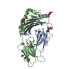
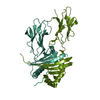
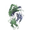
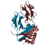
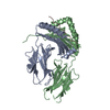
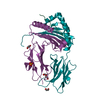
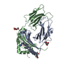
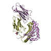
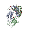
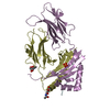
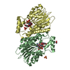
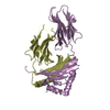
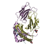
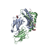
| Title | STRUCTURAL BASIS FOR THE BINDING OF AN IMMUNODOMINANT PEPTIDE FROM MYELIN BASIC PROTEIN IN DIFFERENT REGISTERS BY TWO HLA-DR2 ALLELES | ||||||
 Components Components |
| ||||||
 Keywords Keywords | IMMUNE SYSTEM / MHC class II DR2a | ||||||
| Function / homology |  Function and homology information Function and homology informationstructural constituent of myelin sheath / compact myelin / internode region of axon / myelin assembly / axon ensheathment / autolysosome membrane / myeloid dendritic cell antigen processing and presentation / antigen processing and presentation of endogenous peptide antigen via MHC class II / regulation of T-helper cell differentiation / positive regulation of CD4-positive, CD25-positive, alpha-beta regulatory T cell differentiation ...structural constituent of myelin sheath / compact myelin / internode region of axon / myelin assembly / axon ensheathment / autolysosome membrane / myeloid dendritic cell antigen processing and presentation / antigen processing and presentation of endogenous peptide antigen via MHC class II / regulation of T-helper cell differentiation / positive regulation of CD4-positive, CD25-positive, alpha-beta regulatory T cell differentiation / positive regulation of CD4-positive, alpha-beta T cell activation / EGR2 and SOX10-mediated initiation of Schwann cell myelination / antigen processing and presentation of peptide or polysaccharide antigen via MHC class II / membrane organization / positive regulation of memory T cell differentiation / transport vesicle membrane / Translocation of ZAP-70 to Immunological synapse / Phosphorylation of CD3 and TCR zeta chains / polysaccharide binding / Generation of second messenger molecules / Co-inhibition by PD-1 / immunological synapse / T cell receptor binding / myelination / MHC class II antigen presentation / trans-Golgi network membrane / cell periphery / central nervous system development / lumenal side of endoplasmic reticulum membrane / sensory perception of sound / ER to Golgi transport vesicle membrane / clathrin-coated endocytic vesicle membrane / peptide antigen assembly with MHC class II protein complex / MHC class II protein complex / positive regulation of T cell mediated cytotoxicity / antigen processing and presentation of exogenous peptide antigen via MHC class II / positive regulation of immune response / peptide antigen binding / cognition / response to toxic substance / positive regulation of T cell activation / Interferon gamma signaling / endocytic vesicle membrane / MHC class II protein complex binding / myelin sheath / late endosome membrane / Downstream TCR signaling / protease binding / early endosome membrane / chemical synaptic transmission / adaptive immune response / calmodulin binding / lysosome / immune response / Golgi membrane / lysosomal membrane / neuronal cell body / lipid binding / synapse / cell surface / protein-containing complex / extracellular exosome / nucleus / plasma membrane / cytosol Similarity search - Function | ||||||
| Biological species |  Homo sapiens (human) Homo sapiens (human) | ||||||
| Method |  X-RAY DIFFRACTION / SYNCHROTRON / Resolution: 1.9 Å X-RAY DIFFRACTION / SYNCHROTRON / Resolution: 1.9 Å | ||||||
 Authors Authors | Li, H. / Mariuzza, A.R. / Li, Y. / Martin, R. | ||||||
 Citation Citation | Journal: J.Mol.Biol. / Year: 2000 Title: Structural basis for the binding of an immunodominant peptide from myelin basic protein in different registers by two HLA-DR2 proteins. Authors: Li, Y. / Li, H. / Martin, R. / Mariuzza, R.A. | ||||||
| History |
|
- Structure visualization
| Structure viewer | Molecule: MolmilJmol/JSmol |
|---|
- Downloads & links
-Download
| PDBx/mmCIF format | 1fv1.cif.gz | 177.7 KB | Display | PDBx/mmCIF format |
|---|---|---|---|---|
| PDB format | pdb1fv1.ent.gz | 140 KB | Display | PDB format |
| PDBx/mmJSON format | 1fv1.json.gz | Tree view | PDBx/mmJSON format | |
| Others |  Other downloads Other downloads |
-Validation report
| Arichive directory | https://data.pdbj.org/pub/pdb/validation_reports/fv/1fv1ftp://data.pdbj.org/pub/pdb/validation_reports/fv/1fv1 | HTTPS FTP |
|---|
-Related structure data
| Similar structure data |
|---|

-Links
 PDBj
PDBj


- Assembly
| Deposited unit | 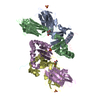
| ||||||||
|---|---|---|---|---|---|---|---|---|---|
| 1 | 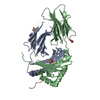
| ||||||||
| 2 | 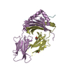
| ||||||||
| Unit cell |
|
-Components
-MAJOR HISTOCOMPATIBILITY COMPLEX ... , 2 types, 4 molecules ADBE
| #1: Protein | Mass: 21084.826 Da / Num. of mol.: 2 Source method: isolated from a genetically manipulated source Source: (gene. exp.) Homo sapiens (human) / Description: HOMO SAPIENS / Production host:  #2: Protein | Mass: 22231.574 Da / Num. of mol.: 2 Source method: isolated from a genetically manipulated source Source: (gene. exp.) Homo sapiens (human) / Description: HOMO SAPIENS / Production host: |
|---|
-Protein/peptide , 1 types, 2 molecules CF
| #3: Protein/peptide | Mass: 2278.627 Da / Num. of mol.: 2 / Fragment: RESIDUES 86-105 / Source method: obtained synthetically Details: The peptide was chemically synthesized. The sequence of the peptide is naturally found in Homo sapiens (human). References: UniProt: P02686 |
|---|
-Non-polymers , 3 types, 499 molecules 


| #4: Chemical |  Mass: 96.063 Da / Num. of mol.: 3 / Source method: obtained synthetically / Formula: SO4 Mass: 96.063 Da / Num. of mol.: 3 / Source method: obtained synthetically / Formula: SO4#5: Chemical |  Mass: 92.094 Da / Num. of mol.: 2 / Source method: obtained synthetically / Formula: C3H8O3 Mass: 92.094 Da / Num. of mol.: 2 / Source method: obtained synthetically / Formula: C3H8O3#6: Water | ChemComp-HOH / | Mass: 18.015 Da / Num. of mol.: 494 / Source method: isolated from a natural source / Formula: H2O |
|---|
-Details
| Has protein modification | Y |
|---|
-Experimental details
-Experiment
| Experiment | Method: X-RAY DIFFRACTION / Number of used crystals: 1 |
|---|
- Sample preparation
| Crystal | Density Matthews: 2.51 Å3/Da / Density % sol: 51.05 % | |||||||||||||||||||||||||
|---|---|---|---|---|---|---|---|---|---|---|---|---|---|---|---|---|---|---|---|---|---|---|---|---|---|---|
| Crystal grow | Temperature: 298 K / Method: evaporation / pH: 6.5 Details: 2/3 dilution of 30% PEG 8000, 0.1M sodium cacodylate, pH6.5, 0.2M ammonium sulfate, EVAPORATION, temperature 298.0K | |||||||||||||||||||||||||
| Crystal grow | *PLUS Method: vapor diffusion, hanging drop | |||||||||||||||||||||||||
| Components of the solutions | *PLUS
|
-Data collection
| Diffraction | Mean temperature: 100 K |
|---|---|
| Diffraction source | Source: SYNCHROTRON / Site: NSLS  / Beamline: X12B / Wavelength: 0.978 / Beamline: X12B / Wavelength: 0.978 |
| Detector | Type: ADSC QUANTUM 4 / Detector: CCD / Date: Nov 7, 1999 |
| Radiation | Protocol: SINGLE WAVELENGTH / Monochromatic (M) / Laue (L): M / Scattering type: x-ray |
| Radiation wavelength | Wavelength: 0.978 Å / Relative weight: 1 |
| Reflection | Resolution: 1.9→100 Å / Num. all: 63997 / Num. obs: 49803 / % possible obs: 0.78 % / Observed criterion σ(F): 2 / Observed criterion σ(I): 1 / Redundancy: 2.8 % / Biso Wilson estimate: 20.5 Å2 / Rmerge(I) obs: 0.081 / Net I/σ(I): 0.081 |
| Reflection shell | Resolution: 1.9→1.99 Å / Redundancy: 2.8 % / Rmerge(I) obs: 0.349 / % possible all: 0.76 |
| Reflection | *PLUS Num. obs: 63997 / % possible obs: 90.8 % / Num. measured all: 181399 |
| Reflection shell | *PLUS % possible obs: 75.6 % / Mean I/σ(I) obs: 2 |
- Processing
| Software |
| ||||||||||||||||||||
|---|---|---|---|---|---|---|---|---|---|---|---|---|---|---|---|---|---|---|---|---|---|
| Refinement | Resolution: 1.9→100 Å / σ(F): 0 / σ(I): 0 / Stereochemistry target values: Engh & Huber
| ||||||||||||||||||||
| Refinement step | Cycle: LAST / Resolution: 1.9→100 Å
| ||||||||||||||||||||
| Refine LS restraints |
| ||||||||||||||||||||
| Software | *PLUS Name: CNS / Classification: refinement | ||||||||||||||||||||
| Refinement | *PLUS Highest resolution: 1.9 Å / Lowest resolution: 100 Å / σ(F): 0 / % reflection Rfree: 5 % | ||||||||||||||||||||
| Solvent computation | *PLUS | ||||||||||||||||||||
| Displacement parameters | *PLUS | ||||||||||||||||||||
| Refine LS restraints | *PLUS Type: c_angle_deg / Dev ideal: 1.5 |
