 Movie
Movie Controller


+ Open data

- Basic information
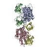
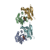
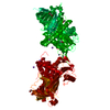
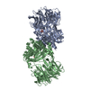
| Entry | Database: PDB / ID: 1iao | ||||||
|---|---|---|---|---|---|---|---|
| Title | CLASS II MHC I-AD IN COMPLEX WITH OVALBUMIN PEPTIDE 323-339 | ||||||
 Components Components | (MHC CLASS II I-AD) x 2 | ||||||
 Keywords Keywords | MHC II / CLASS II MHC / I-A / OVALBUMIN PEPTIDE | ||||||
| Function / homology |  Function and homology information Function and homology informationPhosphorylation of CD3 and TCR zeta chains / Translocation of ZAP-70 to Immunological synapse / Co-inhibition by PD-1 / Generation of second messenger molecules / antigen processing and presentation of peptide antigen / Downstream TCR signaling / MHC class II antigen presentation / positive regulation of T cell differentiation / antigen processing and presentation / multivesicular body ...Phosphorylation of CD3 and TCR zeta chains / Translocation of ZAP-70 to Immunological synapse / Co-inhibition by PD-1 / Generation of second messenger molecules / antigen processing and presentation of peptide antigen / Downstream TCR signaling / MHC class II antigen presentation / positive regulation of T cell differentiation / antigen processing and presentation / multivesicular body / peptide antigen assembly with MHC class II protein complex / MHC class II protein complex / antigen processing and presentation of exogenous peptide antigen via MHC class II / positive regulation of immune response / peptide antigen binding / positive regulation of T cell activation / MHC class II protein complex binding / late endosome membrane / adaptive immune response / early endosome / lysosome / immune response / external side of plasma membrane / lysosomal membrane / protein-containing complex binding / Golgi apparatus / cell surface / membrane / plasma membrane Similarity search - Function | ||||||
| Biological species |  | ||||||
| Method |  X-RAY DIFFRACTION / SYNCHROTRON / MOLECULAR REPLACEMENT / Resolution: 2.6 Å X-RAY DIFFRACTION / SYNCHROTRON / MOLECULAR REPLACEMENT / Resolution: 2.6 Å | ||||||
 Authors Authors | Scott, C.A. / Peterson, P.A. / Teyton, L. / Wilson, I.A. | ||||||
 Citation Citation | Journal: Immunity / Year: 1998 Title: Crystal structures of two I-Ad-peptide complexes reveal that high affinity can be achieved without large anchor residues. Authors: Scott, C.A. / Peterson, P.A. / Teyton, L. / Wilson, I.A. #1: Journal: Immunity / Year: 1998Title: Erratum. Crystal Structures of Two I-Ad-Peptide Complexes Reveal that High Affinity Can be Achieved without Large Anchor Residues Authors: Scott, C.A. / Peterson, P.A. / Teyton, L. / Wilson, I.A. #2: Journal: Protein Sci. / Year: 1998Title: Engineering Protein for X-Ray Crystallography: The Murine Major Histocompatibility Complex Class II Molecule I-Ad Authors: Scott, C.A. / Garcia, K.C. / Stura, E.A. / Peterson, P.A. / Wilson, I.A. / Teyton, L. #3: Journal: J.Exp.Med. / Year: 1996Title: Role of Chain Pairing for the Production of Functional Soluble Ia Major Histocompatibility Complex Class II Molecules Authors: Scott, C.A. / Garcia, K.C. / Carbone, F.R. / Wilson, I.A. / Teyton, L. | ||||||
| History |
|
- Structure visualization
| Structure viewer | Molecule: MolmilJmol/JSmol |
|---|
- Downloads & links
-Download
| PDBx/mmCIF format | 1iao.cif.gz | 90.9 KB | Display | PDBx/mmCIF format |
|---|---|---|---|---|
| PDB format | pdb1iao.ent.gz | 67.7 KB | Display | PDB format |
| PDBx/mmJSON format | 1iao.json.gz | Tree view | PDBx/mmJSON format | |
| Others |  Other downloads Other downloads |
-Validation report
| Arichive directory | https://data.pdbj.org/pub/pdb/validation_reports/ia/1iaoftp://data.pdbj.org/pub/pdb/validation_reports/ia/1iao | HTTPS FTP |
|---|
-Related structure data
| Related structure data |  2iadC 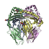 1dlhS S: Starting model for refinement C: citing same article ( |
|---|---|
| Similar structure data |

-Links
 PDBj
PDBj


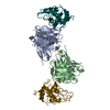
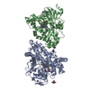
- Assembly
| Deposited unit | 
| ||||||||
|---|---|---|---|---|---|---|---|---|---|
| 1 | 
| ||||||||
| 2 |
| ||||||||
| Unit cell |
|
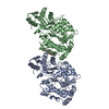
-Components
| #1: Protein | Mass: 22091.631 Da / Num. of mol.: 1 Source method: isolated from a genetically manipulated source Details: BOTH THE A AND B CHAINS HAVE AN 8-RESIDUE (SSADLVPR) PEPTIDE BOUND TO THE C-TERMINUS. THE B CHAIN ALSO HAS A 24-RESIDUE PEPTIDE CONSISTING OF A 2-RESIDUE (RG) SIGNAL SEQUENCE, RESIDUES 323 - ...Details: BOTH THE A AND B CHAINS HAVE AN 8-RESIDUE (SSADLVPR) PEPTIDE BOUND TO THE C-TERMINUS. THE B CHAIN ALSO HAS A 24-RESIDUE PEPTIDE CONSISTING OF A 2-RESIDUE (RG) SIGNAL SEQUENCE, RESIDUES 323 - 339 OF HEN EGG OVALBUMIN AND A 6-RESIDUE (GSGSGS) LINKER, COVALENTLY BONDED TO THE N-TERMINUS. Source: (gene. exp.)  |
|---|---|
| #2: Protein | Mass: 25324.998 Da / Num. of mol.: 1 Source method: isolated from a genetically manipulated source Details: BOTH THE A AND B CHAINS HAVE AN 8-RESIDUE (SSADLVPR) PEPTIDE BOUND TO THE C-TERMINUS. THE B CHAIN ALSO HAS A 24-RESIDUE PEPTIDE CONSISTING OF A 2-RESIDUE (RG) SIGNAL SEQUENCE, RESIDUES 323 - ...Details: BOTH THE A AND B CHAINS HAVE AN 8-RESIDUE (SSADLVPR) PEPTIDE BOUND TO THE C-TERMINUS. THE B CHAIN ALSO HAS A 24-RESIDUE PEPTIDE CONSISTING OF A 2-RESIDUE (RG) SIGNAL SEQUENCE, RESIDUES 323 - 339 OF HEN EGG OVALBUMIN AND A 6-RESIDUE (GSGSGS) LINKER, COVALENTLY BONDED TO THE N-TERMINUS. Source: (gene. exp.) |
| #3: Sugar | ChemComp-NAG /   Type: D-saccharide, beta linking / Mass: 221.208 Da / Num. of mol.: 1 Type: D-saccharide, beta linking / Mass: 221.208 Da / Num. of mol.: 1Source method: isolated from a genetically manipulated source Formula: C8H15NO6 |
| #4: Water | ChemComp-HOH /  Mass: 18.015 Da / Num. of mol.: 8 / Source method: isolated from a natural source / Formula: H2O Mass: 18.015 Da / Num. of mol.: 8 / Source method: isolated from a natural source / Formula: H2O |
| Has protein modification | Y |
-Experimental details
-Experiment
| Experiment | Method: X-RAY DIFFRACTION / Number of used crystals: 1 |
|---|
- Sample preparation
| Crystal | Density Matthews: 2.79 Å3/Da / Density % sol: 56 % | ||||||||||||||||||||
|---|---|---|---|---|---|---|---|---|---|---|---|---|---|---|---|---|---|---|---|---|---|
| Crystal grow | pH: 5.5 / Details: 32% PEG 600, 0.1 IMIDAZOLE MALATE, PH 5.5 | ||||||||||||||||||||
| Crystal | *PLUS | ||||||||||||||||||||
| Crystal grow | *PLUS Temperature: 22 ℃ / Method: vapor diffusion, sitting drop | ||||||||||||||||||||
| Components of the solutions | *PLUS
|
-Data collection
| Diffraction | Mean temperature: 95 K |
|---|---|
| Diffraction source | Source: SYNCHROTRON / Site: SSRL  / Beamline: BL7-1 / Wavelength: 1.08 / Beamline: BL7-1 / Wavelength: 1.08 |
| Detector | Type: MARRESEARCH / Detector: IMAGE PLATE / Date: Apr 1, 1997 Details: 58 CM LONG, PT-COATED, FUSED SILICA, VERTICAL FOCUS |
| Radiation | Monochromator: CYLINDRICALLY BENT TRIANGULAR SI(111) ASYMMETRIC CUT, HORIZONTAL FOCUS Monochromatic (M) / Laue (L): M / Scattering type: x-ray |
| Radiation wavelength | Wavelength: 1.08 Å / Relative weight: 1 |
| Reflection | Resolution: 2.6→27 Å / Num. obs: 14777 / % possible obs: 99 % / Observed criterion σ(I): 0 / Redundancy: 9 % / Rmerge(I) obs: 0.057 / Rsym value: 0.057 / Net I/σ(I): 10 |
| Reflection shell | Resolution: 2.6→2.7 Å / Redundancy: 3.4 % / Rmerge(I) obs: 0.383 / Mean I/σ(I) obs: 2 / Rsym value: 0.383 / % possible all: 91 |
| Reflection shell | *PLUS % possible obs: 91 % / Num. unique obs: 1690 |
- Processing
| Software |
| ||||||||||||||||||||||||||||||||||||||||||||||||||||||||||||
|---|---|---|---|---|---|---|---|---|---|---|---|---|---|---|---|---|---|---|---|---|---|---|---|---|---|---|---|---|---|---|---|---|---|---|---|---|---|---|---|---|---|---|---|---|---|---|---|---|---|---|---|---|---|---|---|---|---|---|---|---|---|
| Refinement | Method to determine structure: MOLECULAR REPLACEMENT Starting model: PDB ENTRY 1DLH Resolution: 2.6→27 Å / Data cutoff high absF: 1000000 / Data cutoff low absF: 0.01 / σ(F): 0
| ||||||||||||||||||||||||||||||||||||||||||||||||||||||||||||
| Displacement parameters | Biso mean: 51 Å2 | ||||||||||||||||||||||||||||||||||||||||||||||||||||||||||||
| Refinement step | Cycle: LAST / Resolution: 2.6→27 Å
| ||||||||||||||||||||||||||||||||||||||||||||||||||||||||||||
| Refine LS restraints |
| ||||||||||||||||||||||||||||||||||||||||||||||||||||||||||||
| LS refinement shell | Resolution: 2.6→2.72 Å / Total num. of bins used: 8
| ||||||||||||||||||||||||||||||||||||||||||||||||||||||||||||
| Software | *PLUS Name: X-PLOR / Version: 3.8 / Classification: refinement | ||||||||||||||||||||||||||||||||||||||||||||||||||||||||||||
| Refinement | *PLUS Rfactor Rfree: 0.32 | ||||||||||||||||||||||||||||||||||||||||||||||||||||||||||||
| Solvent computation | *PLUS | ||||||||||||||||||||||||||||||||||||||||||||||||||||||||||||
| Displacement parameters | *PLUS | ||||||||||||||||||||||||||||||||||||||||||||||||||||||||||||
| Refine LS restraints | *PLUS
|
