 Movie
Movie Controller


+ Open data

- Basic information
| Entry | Database: PDB / ID: 1iak | ||||||
|---|---|---|---|---|---|---|---|
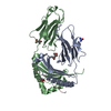
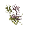
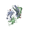
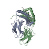
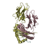
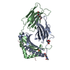
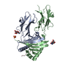
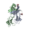
| Title | HISTOCOMPATIBILITY ANTIGEN I-AK | ||||||
 Components Components | (MHC CLASS II I-AK) x 3 | ||||||
 Keywords Keywords | HISTOCOMPATIBILITY ANTIGEN I-AK / HISTOCOMPATIBILITY ANTIGEN / MHC / PEPTIDE COMPLEX | ||||||
| Function / homology |  Function and homology information Function and homology informationPhosphorylation of CD3 and TCR zeta chains / Translocation of ZAP-70 to Immunological synapse / Co-inhibition by PD-1 / Generation of second messenger molecules / antigen processing and presentation of peptide antigen / Downstream TCR signaling / MHC class II antigen presentation / positive regulation of T cell differentiation / antigen processing and presentation / negative regulation of T cell proliferation ...Phosphorylation of CD3 and TCR zeta chains / Translocation of ZAP-70 to Immunological synapse / Co-inhibition by PD-1 / Generation of second messenger molecules / antigen processing and presentation of peptide antigen / Downstream TCR signaling / MHC class II antigen presentation / positive regulation of T cell differentiation / antigen processing and presentation / negative regulation of T cell proliferation / multivesicular body / peptide antigen assembly with MHC class II protein complex / MHC class II protein complex / antigen processing and presentation of exogenous peptide antigen via MHC class II / positive regulation of immune response / peptide antigen binding / positive regulation of T cell activation / cell wall macromolecule catabolic process / lysozyme / lysozyme activity / MHC class II protein complex binding / late endosome membrane / killing of cells of another organism / defense response to Gram-negative bacterium / adaptive immune response / early endosome / lysosome / defense response to Gram-positive bacterium / external side of plasma membrane / lysosomal membrane / protein-containing complex binding / cell surface / Golgi apparatus / extracellular region / plasma membrane / cytoplasm Similarity search - Function | ||||||
| Biological species |  | ||||||
| Method |  X-RAY DIFFRACTION / SYNCHROTRON / MOLECULAR REPLACEMENT / Resolution: 1.9 Å X-RAY DIFFRACTION / SYNCHROTRON / MOLECULAR REPLACEMENT / Resolution: 1.9 Å | ||||||
 Authors Authors | Fremont, D.H. / Unanue, E.R. / Hendrickson, W.A. | ||||||
 Citation Citation | Journal: Immunity / Year: 1998 Title: Crystal structure of I-Ak in complex with a dominant epitope of lysozyme. Authors: Fremont, D.H. / Monnaie, D. / Nelson, C.A. / Hendrickson, W.A. / Unanue, E.R. | ||||||
| History |
|
- Structure visualization
| Structure viewer | Molecule: MolmilJmol/JSmol |
|---|
- Downloads & links
-Download
| PDBx/mmCIF format | 1iak.cif.gz | 101.5 KB | Display | PDBx/mmCIF format |
|---|---|---|---|---|
| PDB format | pdb1iak.ent.gz | 76.5 KB | Display | PDB format |
| PDBx/mmJSON format | 1iak.json.gz | Tree view | PDBx/mmJSON format | |
| Others |  Other downloads Other downloads |
-Validation report
| Arichive directory | https://data.pdbj.org/pub/pdb/validation_reports/ia/1iakftp://data.pdbj.org/pub/pdb/validation_reports/ia/1iak | HTTPS FTP |
|---|
-Related structure data
| Related structure data | 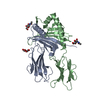 1ieaS S: Starting model for refinement |
|---|---|
| Similar structure data |


-Links
 PDBj
PDBj


- Assembly
| Deposited unit | 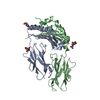
| ||||||||
|---|---|---|---|---|---|---|---|---|---|
| 1 |
| ||||||||
| Unit cell |
| ||||||||
| Components on special symmetry positions |
|
-Components
| #1: Protein | Mass: 22689.281 Da / Num. of mol.: 1 Source method: isolated from a genetically manipulated source Details: WITH COVALENTLY BOUND HEL PEPTIDE / Source: (gene. exp.) | ||||||
|---|---|---|---|---|---|---|---|
| #2: Protein | Mass: 21887.592 Da / Num. of mol.: 1 Source method: isolated from a genetically manipulated source Details: WITH COVALENTLY BOUND HEL PEPTIDE / Source: (gene. exp.) | ||||||
| #3: Protein/peptide | Mass: 1553.697 Da / Num. of mol.: 1 Source method: isolated from a genetically manipulated source Details: WITH COVALENTLY BOUND HEL PEPTIDE / Source: (gene. exp.) | ||||||
| #4: Sugar |   Type: D-saccharide, beta linking / Mass: 221.208 Da / Num. of mol.: 3 Type: D-saccharide, beta linking / Mass: 221.208 Da / Num. of mol.: 3Source method: isolated from a genetically manipulated source Formula: C8H15NO6 #5: Water | ChemComp-HOH / |  Mass: 18.015 Da / Num. of mol.: 369 / Source method: isolated from a natural source / Formula: H2O Mass: 18.015 Da / Num. of mol.: 369 / Source method: isolated from a natural source / Formula: H2OCompound details | THIS ENTRY CONTAINS COORDINATES FOR THE EXTRACELLULAR DOMAINS OF THE MURINE MHC CLASS II MOLECULE I- ...THIS ENTRY CONTAINS COORDINATE | Has protein modification | Y | |
-Experimental details
-Experiment
| Experiment | Method: X-RAY DIFFRACTION / Number of used crystals: 1 |
|---|
- Sample preparation
| Crystal | Density Matthews: 3.4 Å3/Da / Density % sol: 63.85 % | |||||||||||||||||||||||||||||||||||
|---|---|---|---|---|---|---|---|---|---|---|---|---|---|---|---|---|---|---|---|---|---|---|---|---|---|---|---|---|---|---|---|---|---|---|---|---|
| Crystal grow | pH: 4.75 Details: 20% PEG 4000, 7% EG, 80MM A.S, 200MM AMM.ACETATE, 50MM NA.ACETATE PH 4.75 | |||||||||||||||||||||||||||||||||||
| Crystal grow | *PLUS Temperature: 20 ℃ / Method: vapor diffusion, hanging drop | |||||||||||||||||||||||||||||||||||
| Components of the solutions | *PLUS
|
-Data collection
| Diffraction | Mean temperature: 110 K |
|---|---|
| Diffraction source | Source: SYNCHROTRON / Site: NSLS  / Beamline: X4A / Wavelength: 0.9763 / Beamline: X4A / Wavelength: 0.9763 |
| Detector | Type: FUJI / Detector: IMAGE PLATE |
| Radiation | Scattering type: x-ray |
| Radiation wavelength | Wavelength: 0.9763 Å / Relative weight: 1 |
| Reflection | Highest resolution: 1.9 Å / Num. obs: 48139 / % possible obs: 95.7 % / Observed criterion σ(I): 0 / Redundancy: 4.12 % / Biso Wilson estimate: 25.1 Å2 / Rsym value: 0.05 / Net I/σ(I): 11.7 |
| Reflection shell | Resolution: 1.9→1.99 Å / Mean I/σ(I) obs: 2.5 / Rsym value: 0.27 / % possible all: 96.6 |
| Reflection | *PLUS Num. measured all: 198306 / Rmerge(I) obs: 0.05 |
| Reflection shell | *PLUS % possible obs: 96.6 % / Rmerge(I) obs: 0.27 |
- Processing
| Software |
| ||||||||||||||||||||||||||||||||||||||||||||||||||||||||||||||||||||||||||||||||
|---|---|---|---|---|---|---|---|---|---|---|---|---|---|---|---|---|---|---|---|---|---|---|---|---|---|---|---|---|---|---|---|---|---|---|---|---|---|---|---|---|---|---|---|---|---|---|---|---|---|---|---|---|---|---|---|---|---|---|---|---|---|---|---|---|---|---|---|---|---|---|---|---|---|---|---|---|---|---|---|---|---|
| Refinement | Method to determine structure: MOLECULAR REPLACEMENT Starting model: PDB ENTRY 1IEA Resolution: 1.9→10 Å / Rfactor Rfree error: 0.005 / Data cutoff high absF: 100000 / Data cutoff low absF: 0.01 / Isotropic thermal model: RESTRAINED / Cross valid method: THROUGHOUT / σ(F): 0
| ||||||||||||||||||||||||||||||||||||||||||||||||||||||||||||||||||||||||||||||||
| Displacement parameters | Biso mean: 30.9 Å2
| ||||||||||||||||||||||||||||||||||||||||||||||||||||||||||||||||||||||||||||||||
| Refine analyze |
| ||||||||||||||||||||||||||||||||||||||||||||||||||||||||||||||||||||||||||||||||
| Refinement step | Cycle: LAST / Resolution: 1.9→10 Å
| ||||||||||||||||||||||||||||||||||||||||||||||||||||||||||||||||||||||||||||||||
| Refine LS restraints |
| ||||||||||||||||||||||||||||||||||||||||||||||||||||||||||||||||||||||||||||||||
| LS refinement shell | Resolution: 1.9→1.99 Å / Rfactor Rfree error: 0.023 / Total num. of bins used: 8
| ||||||||||||||||||||||||||||||||||||||||||||||||||||||||||||||||||||||||||||||||
| Xplor file |
| ||||||||||||||||||||||||||||||||||||||||||||||||||||||||||||||||||||||||||||||||
| Software | *PLUS Name: X-PLOR / Classification: refinement | ||||||||||||||||||||||||||||||||||||||||||||||||||||||||||||||||||||||||||||||||
| Refine LS restraints | *PLUS
| ||||||||||||||||||||||||||||||||||||||||||||||||||||||||||||||||||||||||||||||||
| LS refinement shell | *PLUS Rfactor obs: 0.357 |
