 Movie
Movie Controller


+ Open data

- Basic information
| Entry | Database: PDB / ID: 1k6q | ||||||
|---|---|---|---|---|---|---|---|
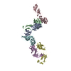
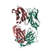
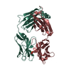
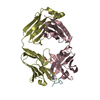
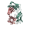
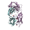
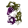
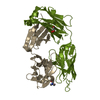
| Title | Crystal structure of antibody Fab fragment D3 | ||||||
 Components Components |
| ||||||
 Keywords Keywords | IMMUNE SYSTEM / antigen-antibody recognition / blood coagulation / tissue factor | ||||||
| Function / homology | Immunoglobulins / Immunoglobulin-like / Sandwich / Mainly Beta / :  Function and homology information Function and homology information | ||||||
| Biological species |  | ||||||
| Method |  X-RAY DIFFRACTION / SYNCHROTRON / MOLECULAR REPLACEMENT / Resolution: 2.4 Å X-RAY DIFFRACTION / SYNCHROTRON / MOLECULAR REPLACEMENT / Resolution: 2.4 Å | ||||||
 Authors Authors | Faelber, K. / Kelley, R.F. / Kirchhofer, D. / Muller, Y.A. | ||||||
 Citation Citation | Journal: To be Published Title: The crystal structure of murine Fab D3 at 2.4 A resolution in comparison with the humanised Fab D3h44 (1.85A) provides structural insight into the humanisation process of the D3 anti-tissue factor antibody Authors: Faelber, K. / Kelley, R.F. / Kirchhofer, D. / Muller, Y.A. #1: Journal: THROMB.HAEMOST. / Year: 2000Title: Epitope location on tissue factor determines the anticoagulant potency of monoclonal anti-tissue factor antibodies Authors: Kirchhofer, D. / Moran, P. / Chiang, N. / Kim, J. / Riederer, M.A. / Eigenbrot, C. / Kelley, R.F. | ||||||
| History |
|
- Structure visualization
| Structure viewer | Molecule: MolmilJmol/JSmol |
|---|
- Downloads & links
-Download
| PDBx/mmCIF format | 1k6q.cif.gz | 96.1 KB | Display | PDBx/mmCIF format |
|---|---|---|---|---|
| PDB format | pdb1k6q.ent.gz | 73.1 KB | Display | PDB format |
| PDBx/mmJSON format | 1k6q.json.gz | Tree view | PDBx/mmJSON format | |
| Others |  Other downloads Other downloads |
-Validation report
| Arichive directory | https://data.pdbj.org/pub/pdb/validation_reports/k6/1k6qftp://data.pdbj.org/pub/pdb/validation_reports/k6/1k6q | HTTPS FTP |
|---|
-Related structure data
| Related structure data | |
|---|---|
| Similar structure data |


-Links
 PDBj
PDBj


- Assembly
| Deposited unit | 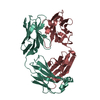
| ||||||||
|---|---|---|---|---|---|---|---|---|---|
| 1 |
| ||||||||
| Unit cell |
|
-Components
| #1: Antibody | Mass: 23121.465 Da / Num. of mol.: 1 / Fragment: Fab fragment Source method: isolated from a genetically manipulated source Source: (gene. exp.)  |
|---|---|
| #2: Antibody | Mass: 23325.008 Da / Num. of mol.: 1 / Fragment: Fab fragment Source method: isolated from a genetically manipulated source Source: (gene. exp.) |
| #3: Water | ChemComp-HOH /  Mass: 18.015 Da / Num. of mol.: 175 / Source method: isolated from a natural source / Formula: H2O Mass: 18.015 Da / Num. of mol.: 175 / Source method: isolated from a natural source / Formula: H2O |
| Has protein modification | Y |
-Experimental details
-Experiment
| Experiment | Method: X-RAY DIFFRACTION / Number of used crystals: 1 |
|---|
- Sample preparation
| Crystal | Density Matthews: 2.4 Å3/Da / Density % sol: 48.66 % |
|---|---|
| Crystal grow | Temperature: 293 K / Method: vapor diffusion, sitting drop / pH: 3.6 Details: lithium sulphate, sodium acetate, pH 3.6, VAPOR DIFFUSION, SITTING DROP, temperature 293K |
-Data collection
| Diffraction | Mean temperature: 110 K |
|---|---|
| Diffraction source | Source: SYNCHROTRON / Site: EMBL/DESY, HAMBURG  / Beamline: BW7B / Wavelength: 0.835 Å / Beamline: BW7B / Wavelength: 0.835 Å |
| Detector | Type: MARRESEARCH / Detector: IMAGE PLATE / Date: Dec 2, 1998 / Details: mirrors |
| Radiation | Monochromator: triangular monochromator / Protocol: SINGLE WAVELENGTH / Monochromatic (M) / Laue (L): M / Scattering type: x-ray |
| Radiation wavelength | Wavelength: 0.835 Å / Relative weight: 1 |
| Reflection | Resolution: 2.39→40 Å / Num. all: 16837 / Num. obs: 16837 / % possible obs: 94.4 % / Observed criterion σ(F): 0 / Observed criterion σ(I): 0 / Redundancy: 3.4 % / Biso Wilson estimate: 40.9 Å2 / Rsym value: 0.077 / Net I/σ(I): 11.7 |
| Reflection shell | Resolution: 2.39→2.58 Å / Redundancy: 3.5 % / Mean I/σ(I) obs: 4.4 / Num. unique all: 3144 / Rsym value: 0.239 / % possible all: 84.5 |
- Processing
| Software |
| |||||||||||||||||||||||||
|---|---|---|---|---|---|---|---|---|---|---|---|---|---|---|---|---|---|---|---|---|---|---|---|---|---|---|
| Refinement | Method to determine structure: MOLECULAR REPLACEMENT Starting model: humanised Fab D3h44 Resolution: 2.4→19.6 Å / Cross valid method: THROUGHOUT / σ(F): 0 / σ(I): 0 / Stereochemistry target values: Engh & Huber
| |||||||||||||||||||||||||
| Displacement parameters | Biso mean: 51.4 Å2
| |||||||||||||||||||||||||
| Refinement step | Cycle: LAST / Resolution: 2.4→19.6 Å
| |||||||||||||||||||||||||
| Refine LS restraints |
| |||||||||||||||||||||||||
| LS refinement shell | Resolution: 2.4→2.55 Å / Rfactor Rfree error: 0.029
|
