 Movie
Movie Controller


+ Open data

- Basic information
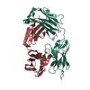
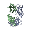
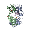
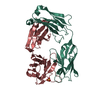
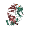
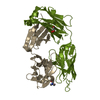
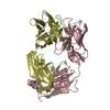
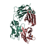
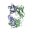
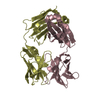
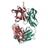
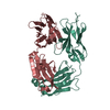
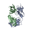
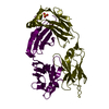
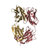
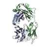
| Entry | Database: PDB / ID: 1jpt | ||||||
|---|---|---|---|---|---|---|---|
| Title | Crystal Structure of Fab D3H44 | ||||||
 Components Components |
| ||||||
 Keywords Keywords | IMMUNE SYSTEM / antigen-antibody recognition / humanized antibody / blood coagulation | ||||||
| Function / homology | Immunoglobulins / Immunoglobulin-like / Sandwich / Mainly Beta Function and homology information Function and homology information | ||||||
| Biological species |  Homo sapiens (human) Homo sapiens (human) | ||||||
| Method |  X-RAY DIFFRACTION / SYNCHROTRON / MOLECULAR REPLACEMENT / Resolution: 1.85 Å X-RAY DIFFRACTION / SYNCHROTRON / MOLECULAR REPLACEMENT / Resolution: 1.85 Å | ||||||
 Authors Authors | Faelber, K. / Kirchhofer, D. / Presta, L. / Kelley, R.F. / Muller, Y.A. | ||||||
 Citation Citation | Journal: J.Mol.Biol. / Year: 2001 Title: The 1.85 A resolution crystal structures of tissue factor in complex with humanized Fab D3h44 and of free humanized Fab D3h44: revisiting the solvation of antigen combining sites. Authors: Faelber, K. / Kirchhofer, D. / Presta, L. / Kelley, R.F. / Muller, Y.A. #1: Journal: THROMB.HAEMOST. / Year: 2001Title: Generation of a humanized, high affinity anti-tissue factor antibody for use as a novel antithrombotic therapeutic Authors: Presta, L. / Sims, P. / Meng, Y.G. / Moran, P. / Bullens, S. / Bunting, S. / Schoenfeld, J. / Lowe, D. / Lai, J. / Rancatore, P. / Iverson, M. / Lim, A. / Chisholm, V. / Kelley, R.F. / ...Authors: Presta, L. / Sims, P. / Meng, Y.G. / Moran, P. / Bullens, S. / Bunting, S. / Schoenfeld, J. / Lowe, D. / Lai, J. / Rancatore, P. / Iverson, M. / Lim, A. / Chisholm, V. / Kelley, R.F. / Riederer, M. / Kirchhofer, D. | ||||||
| History |
|
- Structure visualization
| Structure viewer | Molecule: MolmilJmol/JSmol |
|---|
- Downloads & links
-Download
| PDBx/mmCIF format | 1jpt.cif.gz | 101.4 KB | Display | PDBx/mmCIF format |
|---|---|---|---|---|
| PDB format | pdb1jpt.ent.gz | 76.2 KB | Display | PDB format |
| PDBx/mmJSON format | 1jpt.json.gz | Tree view | PDBx/mmJSON format | |
| Others |  Other downloads Other downloads |
-Validation report
| Arichive directory | https://data.pdbj.org/pub/pdb/validation_reports/jp/1jptftp://data.pdbj.org/pub/pdb/validation_reports/jp/1jpt | HTTPS FTP |
|---|
-Related structure data


-Links
 PDBj
PDBj

- Assembly
| Deposited unit | 
| ||||||||
|---|---|---|---|---|---|---|---|---|---|
| 1 |
| ||||||||
| Unit cell |
|
-Components
| #1: Antibody | Mass: 23523.107 Da / Num. of mol.: 1 / Fragment: Fab fragment Source method: isolated from a genetically manipulated source Source: (gene. exp.) Homo sapiens (human) / Plasmid: pEMX1 / Production host:  |
|---|---|
| #2: Antibody | Mass: 24043.805 Da / Num. of mol.: 1 / Fragment: Fab fragment Source method: isolated from a genetically manipulated source Source: (gene. exp.) Homo sapiens (human) / Plasmid: pEMX1 / Production host: |
| #3: Water | ChemComp-HOH /  Mass: 18.015 Da / Num. of mol.: 372 / Source method: isolated from a natural source / Formula: H2O Mass: 18.015 Da / Num. of mol.: 372 / Source method: isolated from a natural source / Formula: H2O |
| Has protein modification | Y |
-Experimental details
-Experiment
| Experiment | Method: X-RAY DIFFRACTION / Number of used crystals: 1 |
|---|
- Sample preparation
| Crystal | Density Matthews: 2.27 Å3/Da / Density % sol: 45.73 % | ||||||||||||||||||||||||||||||||||||||||||
|---|---|---|---|---|---|---|---|---|---|---|---|---|---|---|---|---|---|---|---|---|---|---|---|---|---|---|---|---|---|---|---|---|---|---|---|---|---|---|---|---|---|---|---|
| Crystal grow | Temperature: 293 K / Method: vapor diffusion, hanging drop / pH: 4.6 Details: ammonium sulphate, sodium acetate, pH 4.6, VAPOR DIFFUSION, HANGING DROP, temperature 293K | ||||||||||||||||||||||||||||||||||||||||||
| Crystal grow | *PLUS Temperature: 20 ℃ / pH: 7.5 / Method: vapor diffusion | ||||||||||||||||||||||||||||||||||||||||||
| Components of the solutions | *PLUS
|
-Data collection
| Diffraction | Mean temperature: 110 K |
|---|---|
| Diffraction source | Source: SYNCHROTRON / Site: ESRF  / Beamline: BM14 / Wavelength: 0.933 Å / Beamline: BM14 / Wavelength: 0.933 Å |
| Detector | Type: MARRESEARCH / Detector: CCD / Date: Dec 10, 1999 / Details: mirrors |
| Radiation | Monochromator: diamond + germanium / Protocol: SINGLE WAVELENGTH / Monochromatic (M) / Laue (L): M / Scattering type: x-ray |
| Radiation wavelength | Wavelength: 0.933 Å / Relative weight: 1 |
| Reflection | Resolution: 1.85→40 Å / Num. all: 36063 / Num. obs: 36063 / % possible obs: 96.3 % / Observed criterion σ(F): 0 / Observed criterion σ(I): 0 / Redundancy: 3.4 % / Biso Wilson estimate: 24.7 Å2 / Rsym value: 0.077 / Net I/σ(I): 11.3 |
| Reflection shell | Resolution: 1.85→1.9 Å / Redundancy: 3.4 % / Mean I/σ(I) obs: 2.8 / Num. unique all: 2779 / Rsym value: 0.416 / % possible all: 91.6 |
| Reflection | *PLUS Lowest resolution: 40 Å / Num. measured all: 124061 / Rmerge(I) obs: 0.077 |
| Reflection shell | *PLUS % possible obs: 91.6 % / Rmerge(I) obs: 0.416 |
- Processing
| Software |
| |||||||||||||||||||||||||
|---|---|---|---|---|---|---|---|---|---|---|---|---|---|---|---|---|---|---|---|---|---|---|---|---|---|---|
| Refinement | Method to determine structure: MOLECULAR REPLACEMENT Starting model: complex D3h44 - TF Resolution: 1.85→40 Å / Cross valid method: THROUGHOUT / σ(F): 0 / σ(I): 0 / Stereochemistry target values: Engh & Huber
| |||||||||||||||||||||||||
| Displacement parameters | Biso mean: 20.5 Å2
| |||||||||||||||||||||||||
| Refinement step | Cycle: LAST / Resolution: 1.85→40 Å
| |||||||||||||||||||||||||
| Refine LS restraints |
| |||||||||||||||||||||||||
| LS refinement shell | Resolution: 1.85→1.97 Å / Rfactor Rfree error: 0.013
| |||||||||||||||||||||||||
| Software | *PLUS Name: CNS / Version: 1 / Classification: refinement | |||||||||||||||||||||||||
| Refinement | *PLUS Lowest resolution: 40 Å / Num. reflection obs: 33469 / σ(F): 0 / Num. reflection Rfree: 2550 / Rfactor obs: 0.183 | |||||||||||||||||||||||||
| Solvent computation | *PLUS | |||||||||||||||||||||||||
| Displacement parameters | *PLUS Biso mean: 20.5 Å2 | |||||||||||||||||||||||||
| Refine LS restraints | *PLUS Type: c_angle_deg / Dev ideal: 1.79 | |||||||||||||||||||||||||
| LS refinement shell | *PLUS Rfactor Rfree: 0.27 / Rfactor Rwork: 0.242 |
