 Movie
Movie Controller


+ Open data

- Basic information
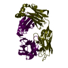
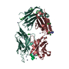
| Entry | Database: PDB / ID: 2q76 | ||||||
|---|---|---|---|---|---|---|---|
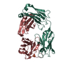
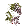
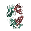
| Title | Mouse anti-hen egg white lysozyme antibody F10.6.6 Fab fragment | ||||||
 Components Components |
| ||||||
 Keywords Keywords | IMMUNE SYSTEM / Antibody / Fab fragment / lysozyme / affinity maturation / VH-VL interface. | ||||||
| Function / homology |  Function and homology information Function and homology informationalpha-beta T cell receptor complex / IgG immunoglobulin complex / B cell differentiation / adaptive immune response / extracellular region / plasma membrane Similarity search - Function | ||||||
| Biological species |  | ||||||
| Method |  X-RAY DIFFRACTION / SYNCHROTRON / MOLECULAR REPLACEMENT / Resolution: 2 Å X-RAY DIFFRACTION / SYNCHROTRON / MOLECULAR REPLACEMENT / Resolution: 2 Å | ||||||
 Authors Authors | Cauerhff, A. / Klinke, S. / Acierno, J.P. / Goldbaum, F.A. / Braden, B.C. | ||||||
 Citation Citation | Journal: J.Mol.Biol. / Year: 2007 Title: Affinity maturation increases the stability and plasticity of the Fv domain of anti-protein antibodies. Authors: Acierno, J.P. / Braden, B.C. / Klinke, S. / Goldbaum, F.A. / Cauerhff, A. #1: Journal: Proc.Natl.Acad.Sci.USA / Year: 2004Title: Structural mechanism for affinity maturation of an anti-lysozyme antibody Authors: Cauerhff, A. / Goldbaum, F.A. / Braden, B.C. | ||||||
| History |
| ||||||
| Remark 999 | sequence The sequences for Fab F10.6.6 fragment Light and heavy chains are not in the UNP database ...sequence The sequences for Fab F10.6.6 fragment Light and heavy chains are not in the UNP database at the time of processing. Author indicated the sequence references AF110316.3 GI:32456055 for VH, and AY277254.1 GI:33642144 for VL, respectively. Both sequences are identical to the sequences in the coordinates. |
- Structure visualization
| Structure viewer | Molecule: MolmilJmol/JSmol |
|---|
- Downloads & links
-Download
| PDBx/mmCIF format | 2q76.cif.gz | 181 KB | Display | PDBx/mmCIF format |
|---|---|---|---|---|
| PDB format | pdb2q76.ent.gz | 144.9 KB | Display | PDB format |
| PDBx/mmJSON format | 2q76.json.gz | Tree view | PDBx/mmJSON format | |
| Others |  Other downloads Other downloads |
-Validation report
| Arichive directory | https://data.pdbj.org/pub/pdb/validation_reports/q7/2q76ftp://data.pdbj.org/pub/pdb/validation_reports/q7/2q76 | HTTPS FTP |
|---|
-Related structure data
| Related structure data | 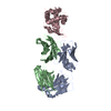 1p2cS S: Starting model for refinement |
|---|---|
| Similar structure data |


-Links
 PDBj
PDBj


- Assembly
| Deposited unit | 
| ||||||||
|---|---|---|---|---|---|---|---|---|---|
| 1 | 
| ||||||||
| 2 | 
| ||||||||
| Unit cell |
|
-Components
| #1: Antibody | Mass: 23320.539 Da / Num. of mol.: 2 / Fragment: domain VL and CL / Source method: isolated from a natural source Details: The antibody was obtained from ascitic fluid generated by injecting monoclonal hybridoma cells. Source: (natural) #2: Antibody | Mass: 23198.896 Da / Num. of mol.: 2 / Fragment: domain VH and CH1 / Source method: isolated from a natural source Details: The antibody was obtained from ascitic fluid generated by injecting monoclonal hybridoma cells. Source: (natural) #3: Water | ChemComp-HOH / |  Mass: 18.015 Da / Num. of mol.: 517 / Source method: isolated from a natural source / Formula: H2O Mass: 18.015 Da / Num. of mol.: 517 / Source method: isolated from a natural source / Formula: H2OHas protein modification | Y | |
|---|
-Experimental details
-Experiment
| Experiment | Method: X-RAY DIFFRACTION / Number of used crystals: 1 |
|---|
- Sample preparation
| Crystal | Density Matthews: 2.14 Å3/Da / Density % sol: 42.63 % |
|---|---|
| Crystal grow | Temperature: 298 K / Method: vapor diffusion, hanging drop / pH: 4.7 Details: The F10.6.6 Fab fragment was crystallized in a mother liquor containing 24% (w/v) PEG 1000, 0.2 M CaCl2 and 0.1 M acetate buffer pH 4.7. Large prisms of about 0.30 mm x 0.05 mm x 0.05 mm ...Details: The F10.6.6 Fab fragment was crystallized in a mother liquor containing 24% (w/v) PEG 1000, 0.2 M CaCl2 and 0.1 M acetate buffer pH 4.7. Large prisms of about 0.30 mm x 0.05 mm x 0.05 mm were obtained after several weeks. VAPOR DIFFUSION, HANGING DROP, temperature 298K, pH 4.70 |
-Data collection
| Diffraction | Mean temperature: 100 K |
|---|---|
| Diffraction source | Source: SYNCHROTRON / Site: LNLS  / Beamline: D03B-MX1 / Wavelength: 1.431 Å / Beamline: D03B-MX1 / Wavelength: 1.431 Å |
| Detector | Type: MAR CCD 165 mm / Detector: CCD / Date: May 14, 2003 |
| Radiation | Monochromator: SI SINGLE CRYSTAL / Protocol: SINGLE WAVELENGTH / Monochromatic (M) / Laue (L): M / Scattering type: x-ray |
| Radiation wavelength | Wavelength: 1.431 Å / Relative weight: 1 |
| Reflection | Resolution: 2→50 Å / Num. all: 50300 / Num. obs: 50300 / % possible obs: 96 % / Observed criterion σ(F): 0 / Observed criterion σ(I): 0 / Redundancy: 8.1 % / Biso Wilson estimate: 19.3 Å2 / Rmerge(I) obs: 0.069 / Rsym value: 0.069 / Net I/σ(I): 9.8 |
| Reflection shell | Resolution: 2→2.07 Å / Redundancy: 8.2 % / Rmerge(I) obs: 0.195 / Mean I/σ(I) obs: 3.3 / Num. unique all: 7122 / Rsym value: 0.195 / % possible all: 92 |
- Processing
| Software |
| |||||||||||||||
|---|---|---|---|---|---|---|---|---|---|---|---|---|---|---|---|---|
| Refinement | Method to determine structure: MOLECULAR REPLACEMENT Starting model: Fab fragment of PDB 1P2C Resolution: 2→50 Å / Cross valid method: THROUGHOUT / σ(F): 2 / Stereochemistry target values: Engh & Huber
| |||||||||||||||
| Displacement parameters | Biso mean: 22 Å2 | |||||||||||||||
| Refinement step | Cycle: LAST / Resolution: 2→50 Å
| |||||||||||||||
| Refine LS restraints |
|
