 Movie
Movie Controller


+ Open data

- Basic information
| Entry | Database: PDB / ID: 1200000000 | ||||||
|---|---|---|---|---|---|---|---|
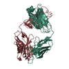
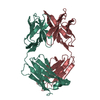
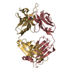
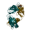
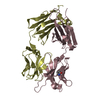
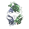
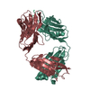
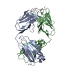
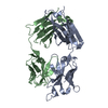
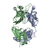
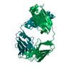
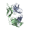
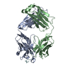
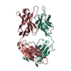
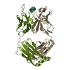
| Title | 2E8 FAB FRAGMENT | ||||||
 Components Components |
| ||||||
 Keywords Keywords | IMMUNOGLOBULIN | ||||||
| Function / homology | Immunoglobulins / Immunoglobulin-like / Sandwich / Mainly Beta / : / :  Function and homology information Function and homology information | ||||||
| Biological species |  | ||||||
| Method |  X-RAY DIFFRACTION / MOLECULAR REPLACEMENT / Resolution: 1.9 Å X-RAY DIFFRACTION / MOLECULAR REPLACEMENT / Resolution: 1.9 Å | ||||||
 Authors Authors | Rupp, B. / Trakhanov, S. | ||||||
 Citation Citation | Journal: Acta Crystallogr.,Sect.D / Year: 1999 Title: Structure of a monoclonal 2E8 Fab antibody fragment specific for the low-density lipoprotein-receptor binding region of apolipoprotein E refined at 1.9 A. Authors: Trakhanov, S. / Parkin, S. / Raffai, R. / Milne, R. / Newhouse, Y.M. / Weisgraber, K.H. / Rupp, B. | ||||||
| History |
|
- Structure visualization
| Structure viewer | Molecule: MolmilJmol/JSmol |
|---|
- Downloads & links
-Download
| PDBx/mmCIF format | 12e8.cif.gz | 193.6 KB | Display | PDBx/mmCIF format |
|---|---|---|---|---|
| PDB format | pdb12e8.ent.gz | 153.1 KB | Display | PDB format |
| PDBx/mmJSON format | 12e8.json.gz | Tree view | PDBx/mmJSON format | |
| Others |  Other downloads Other downloads |
-Validation report
| Arichive directory | https://data.pdbj.org/pub/pdb/validation_reports/2e/12e8ftp://data.pdbj.org/pub/pdb/validation_reports/2e/12e8 | HTTPS FTP |
|---|
-Related structure data


-Links
 PDBj
PDBj

- Assembly
| Deposited unit | 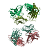
| ||||||||||||||||||||
|---|---|---|---|---|---|---|---|---|---|---|---|---|---|---|---|---|---|---|---|---|---|
| 1 | 
| ||||||||||||||||||||
| 2 | 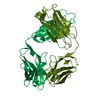
| ||||||||||||||||||||
| Unit cell |
| ||||||||||||||||||||
| Noncrystallographic symmetry (NCS) | NCS oper:
|
-Components
| #1: Antibody | Mass: 23540.990 Da / Num. of mol.: 2 / Fragment: FAB FRAGMENT / Source method: isolated from a natural source / Source: (natural) #2: Antibody | Mass: 23878.586 Da / Num. of mol.: 2 / Fragment: FAB FRAGMENT / Source method: isolated from a natural source / Source: (natural) #3: Water | ChemComp-HOH / |  Mass: 18.015 Da / Num. of mol.: 767 / Source method: isolated from a natural source / Formula: H2O Mass: 18.015 Da / Num. of mol.: 767 / Source method: isolated from a natural source / Formula: H2OHas protein modification | Y | Sequence details | AMINO ACIDS ARE NUMBERED ACCORDING TO THE KABAT NUMBERING SYSTEM FOR ANTIBODIES | |
|---|
-Experimental details
-Experiment
| Experiment | Method: X-RAY DIFFRACTION / Number of used crystals: 2 |
|---|
- Sample preparation
| Crystal | Density Matthews: 2.37 Å3/Da / Density % sol: 48 % | ||||||||||||||||||||||||||||||
|---|---|---|---|---|---|---|---|---|---|---|---|---|---|---|---|---|---|---|---|---|---|---|---|---|---|---|---|---|---|---|---|
| Crystal grow | pH: 5.6 Details: 23% PEG 4000 (W/V), 0.10 M SODIUM CITRATE, AND 0.2 M AMMONIUM ACETATE (PH 5.6). CRYSTALS (0.2 X 0.2 X 0.5 MM) WERE OBTAINED IN 4-6 WEEKS. | ||||||||||||||||||||||||||||||
| Crystal | *PLUS | ||||||||||||||||||||||||||||||
| Crystal grow | *PLUS Method: vapor diffusion, hanging drop | ||||||||||||||||||||||||||||||
| Components of the solutions | *PLUS
|
-Data collection
| Diffraction | Mean temperature: 125 K |
|---|---|
| Diffraction source | Source: ROTATING ANODE / Type: RIGAKU RUH2R / Wavelength: 1.5418 |
| Detector | Type: ADSC / Detector: AREA DETECTOR / Date: May 1, 1996 / Details: COLLIMATOR |
| Radiation | Monochromator: GRAPHITE(002) / Monochromatic (M) / Laue (L): M / Scattering type: x-ray |
| Radiation wavelength | Wavelength: 1.5418 Å / Relative weight: 1 |
| Reflection | Highest resolution: 1.85 Å / Num. obs: 63078 / % possible obs: 84 % / Observed criterion σ(I): 0 / Redundancy: 3.8 % / Biso Wilson estimate: 11.5 Å2 / Rmerge(I) obs: 0.085 / Rsym value: 0.071 / Net I/σ(I): 6 |
| Reflection shell | Resolution: 1.85→2 Å / Redundancy: 2.1 % / Rmerge(I) obs: 0.39 / Mean I/σ(I) obs: 1.6 / Rsym value: 0.27 / % possible all: 82 |
| Reflection | *PLUS Num. measured all: 237795 |
- Processing
| Software |
| ||||||||||||||||||||||||||||||||||||||||||||||||||||||||||||||||||||||||||||||||
|---|---|---|---|---|---|---|---|---|---|---|---|---|---|---|---|---|---|---|---|---|---|---|---|---|---|---|---|---|---|---|---|---|---|---|---|---|---|---|---|---|---|---|---|---|---|---|---|---|---|---|---|---|---|---|---|---|---|---|---|---|---|---|---|---|---|---|---|---|---|---|---|---|---|---|---|---|---|---|---|---|---|
| Refinement | Method to determine structure: MOLECULAR REPLACEMENT Starting model: PDB ENTRIES 6FAB AND 1FLR Resolution: 1.9→20 Å / Rfactor Rfree error: 0.008 / Data cutoff high absF: 10000000 / Data cutoff low absF: 0.001 / Isotropic thermal model: RESTRAINED / Cross valid method: A POSTERIORI / σ(F): 0 Details: BULK SOLVENT MODEL USED. THE CONNECTING LOOP BETWEEN BETA STRANDS B AND C IN THE CH1 DOMAIN IS POORLY DEFINED IN THE STRUCTURE. ATTEMPTS TO IMPROVE THE LOOP H123-H133 USING VARIOUS NCS ...Details: BULK SOLVENT MODEL USED. THE CONNECTING LOOP BETWEEN BETA STRANDS B AND C IN THE CH1 DOMAIN IS POORLY DEFINED IN THE STRUCTURE. ATTEMPTS TO IMPROVE THE LOOP H123-H133 USING VARIOUS NCS AVERAGING SCHEMES AS WELL AS AUTOMATED REBUILDING AND REFINEMENT WITH ARP DID NOT IMPROVE THIS REGION. HOWEVER, USING ARP THE TRACING OF CH BETA STRANDS B-E WAS CONFIRMED INDEPENDENT OF ANY STARTING MODEL. THE CONNECTING LOOP BETWEEN BETA STRANDS B AND C IN THE CH1 DOMAIN IS POORLY DEFINED IN THE STRUCTURE. ATTEMPTS TO IMPROVE THE LOOP H123-H133 USING VARIOUS NCS AVERAGING SCHEMES AS WELL AS AUTOMATED REBUILDING AND REFINEMENT WITH ARP DID NOT IMPROVE THIS REGION. HOWEVER, USING ARP THE TRACING OF CH BETA STRANDS B-E WAS CONFIRMED INDEPENDENT OF ANY STARTING MODEL.
| ||||||||||||||||||||||||||||||||||||||||||||||||||||||||||||||||||||||||||||||||
| Displacement parameters | Biso mean: 16.9 Å2 | ||||||||||||||||||||||||||||||||||||||||||||||||||||||||||||||||||||||||||||||||
| Refine analyze |
| ||||||||||||||||||||||||||||||||||||||||||||||||||||||||||||||||||||||||||||||||
| Refinement step | Cycle: LAST / Resolution: 1.9→20 Å
| ||||||||||||||||||||||||||||||||||||||||||||||||||||||||||||||||||||||||||||||||
| Refine LS restraints |
| ||||||||||||||||||||||||||||||||||||||||||||||||||||||||||||||||||||||||||||||||
| LS refinement shell | Resolution: 1.9→2.02 Å / Rfactor Rfree error: 0.026 / Total num. of bins used: 6
| ||||||||||||||||||||||||||||||||||||||||||||||||||||||||||||||||||||||||||||||||
| Xplor file |
| ||||||||||||||||||||||||||||||||||||||||||||||||||||||||||||||||||||||||||||||||
| Software | *PLUS Name: X-PLOR / Version: 3.851 / Classification: refinement | ||||||||||||||||||||||||||||||||||||||||||||||||||||||||||||||||||||||||||||||||
| Refine LS restraints | *PLUS
|
